- Bioactive Compounds
- By Signaling Pathways
- PI3K/Akt/mTOR
- Epigenetics
- Methylation
- Immunology & Inflammation
- Protein Tyrosine Kinase
- Angiogenesis
- Apoptosis
- Autophagy
- ER stress & UPR
- JAK/STAT
- MAPK
- Cytoskeletal Signaling
- Cell Cycle
- TGF-beta/Smad
- Compound Libraries
- Popular Compound Libraries
- Customize Library
- Clinical and FDA-approved Related
- Bioactive Compound Libraries
- Inhibitor Related
- Natural Product Related
- Metabolism Related
- Cell Death Related
- By Signaling Pathway
- By Disease
- Anti-infection and Antiviral Related
- Neuronal and Immunology Related
- Fragment and Covalent Related
- FDA-approved Drug Library
- FDA-approved & Passed Phase I Drug Library
- Preclinical/Clinical Compound Library
- Bioactive Compound Library-I
- Bioactive Compound Library-II
- Kinase Inhibitor Library
- Express-Pick Library
- Natural Product Library
- Human Endogenous Metabolite Compound Library
- Alkaloid Compound LibraryNew
- Angiogenesis Related compound Library
- Anti-Aging Compound Library
- Anti-alzheimer Disease Compound Library
- Antibiotics compound Library
- Anti-cancer Compound Library
- Anti-cancer Compound Library-Ⅱ
- Anti-cancer Metabolism Compound Library
- Anti-Cardiovascular Disease Compound Library
- Anti-diabetic Compound Library
- Anti-infection Compound Library
- Antioxidant Compound Library
- Anti-parasitic Compound Library
- Antiviral Compound Library
- Apoptosis Compound Library
- Autophagy Compound Library
- Calcium Channel Blocker LibraryNew
- Cambridge Cancer Compound Library
- Carbohydrate Metabolism Compound LibraryNew
- Cell Cycle compound library
- CNS-Penetrant Compound Library
- Covalent Inhibitor Library
- Cytokine Inhibitor LibraryNew
- Cytoskeletal Signaling Pathway Compound Library
- DNA Damage/DNA Repair compound Library
- Drug-like Compound Library
- Endoplasmic Reticulum Stress Compound Library
- Epigenetics Compound Library
- Exosome Secretion Related Compound LibraryNew
- FDA-approved Anticancer Drug LibraryNew
- Ferroptosis Compound Library
- Flavonoid Compound Library
- Fragment Library
- Glutamine Metabolism Compound Library
- Glycolysis Compound Library
- GPCR Compound Library
- Gut Microbial Metabolite Library
- HIF-1 Signaling Pathway Compound Library
- Highly Selective Inhibitor Library
- Histone modification compound library
- HTS Library for Drug Discovery
- Human Hormone Related Compound LibraryNew
- Human Transcription Factor Compound LibraryNew
- Immunology/Inflammation Compound Library
- Inhibitor Library
- Ion Channel Ligand Library
- JAK/STAT compound library
- Lipid Metabolism Compound LibraryNew
- Macrocyclic Compound Library
- MAPK Inhibitor Library
- Medicine Food Homology Compound Library
- Metabolism Compound Library
- Methylation Compound Library
- Mouse Metabolite Compound LibraryNew
- Natural Organic Compound Library
- Neuronal Signaling Compound Library
- NF-κB Signaling Compound Library
- Nucleoside Analogue Library
- Obesity Compound Library
- Oxidative Stress Compound LibraryNew
- Plant Extract Library
- Phenotypic Screening Library
- PI3K/Akt Inhibitor Library
- Protease Inhibitor Library
- Protein-protein Interaction Inhibitor Library
- Pyroptosis Compound Library
- Small Molecule Immuno-Oncology Compound Library
- Mitochondria-Targeted Compound LibraryNew
- Stem Cell Differentiation Compound LibraryNew
- Stem Cell Signaling Compound Library
- Natural Phenol Compound LibraryNew
- Natural Terpenoid Compound LibraryNew
- TGF-beta/Smad compound library
- Traditional Chinese Medicine Library
- Tyrosine Kinase Inhibitor Library
- Ubiquitination Compound Library
-
Cherry Picking
You can personalize your library with chemicals from within Selleck's inventory. Build the right library for your research endeavors by choosing from compounds in all of our available libraries.
Please contact us at [email protected] to customize your library.
You could select:
- Antibodies
- Bioreagents
- qPCR
- 2x SYBR Green qPCR Master Mix
- 2x SYBR Green qPCR Master Mix(Low ROX)
- 2x SYBR Green qPCR Master Mix(High ROX)
- Protein Assay
- Protein A/G Magnetic Beads for IP
- Anti-DYKDDDDK Tag magnetic beads
- Anti-DYKDDDDK Tag Affinity Gel
- Anti-Myc magnetic beads
- Anti-HA magnetic beads
- Poly DYKDDDDK Tag Peptide lyophilized powder
- Protease Inhibitor Cocktail
- Protease Inhibitor Cocktail (EDTA-Free, 100X in DMSO)
- Phosphatase Inhibitor Cocktail (2 Tubes, 100X)
- Cell Biology
- Cell Counting Kit-8 (CCK-8)
- Animal Experiment
- Mouse Direct PCR Kit (For Genotyping)
- New Products
- Contact Us
Choose Your Country or Region
-
 Australia
-
 Austria
-
 Belgium
-
 Brazil
-
 Canada
-
 China
China
-
 Czech Republic
-
 Denmark
-
 Finland
-
 France
-
 Germany
-
 Greece
-
 Hong Kong
-
 Hungary
-
 Iceland
-
 India
India
-
 Ireland
-
 Israel
-
 Italy
Italy
-
 Japan
Japan
-
 Korea
Korea
-
 Luxembourg
-
 Malaysia
-
 Netherlands
-
 New Zealand
-
 Norway
-
 Poland
-
 Qatar
Qatar
-
 Romania
-
 Saudi Arabia
-
 Singapore
-
 Spain
Spain
-
 Sweden
-
 Switzerland
-
 Taiwan
-
 Turkey
-
 United Kingdom
-
 United States
-
 Other Countries
Home
Blog of Signal Transduction
Epigenetics
Aurora Kinase
The distribution of primary cilia in the mouse embyo
Category
- PI3K/Akt/mTOR
- Epigenetics
- Methylation
- Immunology & Inflammation
- CD markers
- PD-1/PD-L1
- TNF-alpha
- COX
- CCR
- Histamine Receptor
- IL Receptor
- Anti-infection
- AhR
- IDO/TDO
- gp120/CD4
- NOD
- CXCR
- MALT
- LTR
- TLR
- NOS
- Nrf2
- ROS
- NADPH-oxidase
- Immunology & Inflammation related
- FKBP
- TRIF
- AKR1C
- PKR
- TpoR
- Parasite
- Heme Oxygenase
- MmpL3
- MyD88
- eIF
- Antioxidant
- STING
- MIF
- Galectin
- Nur77
- Complement System
- Prostaglandin Receptor
- SPHK
- Glutathione
- β-lactamase
- PGES
- TBK1
- IFN
- IRAK
- FLAP
- Interleukins
- Arginase
- NLRP3
- Pyroptosis
- cGAS
- Neuraminidase
- SIK
- PGDS
- Protein Tyrosine Kinase
- Angiogenesis
- Apoptosis
- Autophagy
- ER stress & UPR
- JAK/STAT
- MAPK
- Cytoskeletal Signaling
- Cell Cycle
- TGF-beta/Smad
- DNA Damage/DNA Repair
- Stem Cells & Wnt
- Hippo
- Ubiquitin
- Neuronal Signaling
- Calcium Channel
- Beta Amyloid
- 5-HT Receptor
- COX
- GluR
- Adrenergic Receptor
- AChR
- Histamine Receptor
- Dopamine Receptor
- Opioid Receptor
- GABA Receptor
- P-gp
- P2 Receptor
- Cannabinoid Receptor
- OX Receptor
- CGRP Receptor
- MT Receptor
- MAO
- FAAH
- GlyT
- NMDAR
- CaMK
- BACE
- Trk receptor
- NPY receptor
- CCK receptor
- Serotonin Transporter
- COMT
- Neurotensin Receptor
- Melanocortin Receptor
- TRP Channel
- Adenosine Deaminase
- Adenosine Kinase
- BChE
- EAAT
- Neurokinin Receptor
- Notch
- Imidazoline Receptor
- Sigma Receptor
- NF-κB
- GPCR & G Protein
- Ras
- 5-HT Receptor
- CCR
- GluR
- Adrenergic Receptor
- AChR
- Histamine Receptor
- Dopamine Receptor
- Opioid Receptor
- GPR
- P2 Receptor
- Cannabinoid Receptor
- Endothelin Receptor
- S1P Receptor
- Hedgehog/Smoothened
- SGLT
- LPA Receptor
- OX Receptor
- CGRP Receptor
- MT Receptor
- PAFR
- PKA
- Vasopressin Receptor
- cAMP
- CXCR
- TAAR
- CaSR
- Glucagon Receptor
- LTR
- Adenosine Receptor
- Guanylate Cyclase
- GHSR
- CCK receptor
- GRK
- Leukotriene
- FPR
- Prostaglandin Receptor
- TSH Receptor
- PKD
- GNRH Receptor
- Neurokinin Receptor
- CRFR
- PKG
- Bradykinin Receptor
- Angiotensin Receptor
- Imidazoline Receptor
- Bombesin Receptor
- RGS
- Neurotensin Receptor
- Sigma Receptor
- Melanocortin Receptor
- PAR
- Taste Receptor
- NPY receptor
- Parathyroid Hormone Receptor
- DOCK
- AdipoR
- GPCR19
- Endocrinology & Hormones
- Transmembrane Transporters
- Metabolism
- PPAR
- P450 (e.g. CYP17)
- HSP (HSP90)
- PDE
- Hydroxylase
- Factor Xa
- DHFR
- Aminopeptidase
- Dehydrogenase
- Procollagen C Proteinase
- Phospholipase (e.g. PLA)
- DPP
- Carbonic Anhydrase
- Phosphorylase
- Liver X Receptor
- FAAH
- Acyltransferase
- PKM
- Lipoxygenase
- FOX
- Lipase
- Fatty Acid Synthase
- LDL
- NAMPT
- Decarboxylase
- Casein Kinase
- Thioredoxin
- Retinoid Receptor
- FXR
- Transferase
- CETP
- Serine/threonin kinase
- IDO/TDO
- phosphatase
- GLUT
- HMG-CoA Reductase
- Vitamin
- SGK
- GOT
- 3-MST
- GTPCH
- glucocerebrosidase
- FABP
- Serine hydrolase
- Epoxide Hydrolase
- ACSS2
- ROR
- Catalase
- THR
- Thioredoxin Reductase
- PDHK
- Glucosylceramide Synthase
- ACE
- Mannosidase
- PGC-1α
- PARG
- Xanthine Oxidase
- CPSase
- Leukotriene
- Adenosine Deaminase
- Aldose Reductase
- Glutathione
- Acetyl-CoA carboxylase
- Adenosine Kinase
- OXPHOS
- SHIP
- PCSK9
- Mitochondrial Metabolism
- Peroxidases
- PREP
- PGDS
- Mitochondrial pyruvate carrier
- Neprilysin
- PP2A
- FTO
- RUNX
- SREBP
- AdipoR
- PAD
- CPT
- SCD
- Carbohydrate Metabolism
- CAR
- AP-1
- LDH
- γGCS
- SSTR
- PAI-1
- FTase
- SOD
- ATP-citrate lyase
- FAO
- Proteases
- Microbiology
- Others
- ADC Cytotoxin
- ADC Linker
- Drug-Linker Conjugates for ADC
- Antineoplastic and Immunosuppressive Antibiotics
- Selection Antibiotics for Transfected Cell
- Antibiotics for Mammalian Cell Culture
- Antibiotics for Plant Cell Culture
- Hydrotropic Agents
- Dyes
- PROTAC
- PROTAC Linker
- E3 ligase Ligand
- Target Protein Ligand
- Others
- Antibody-Drug Conjugate
Archives
The distribution of primary cilia in the mouse embyo

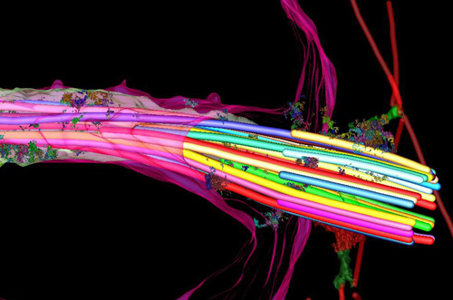
Primary cilia is formed by centrioles project from the surface of most vertebrate cells, and is involved in the response to specific developmental signals, including Hedgehog (Hh) ligands. Bangs et al. demonstrate the time and location that primary cilia appear in the mouse embryo. The article was published on Nature Cell Biology, recently.
Researchers established a transgenic mouse embryo cell line that express ARL13B fused to the red fluorescent protein mCherry in cilia, and Centrin 2 fused to GFP in centrosomes. At E6.0, primary cilia first appear in the epiblast on cells, following by the present on all derivatives of the epiblast. On the other hand, extraembryonic cell lines, such as visceral endoderm and trophectoderm lineages, are absent of cilia but have centrosomes. The results match with what they found in vivo. In addition, extraembryonic endoderm (XEN) stem cells, which lack cilia, can form cilia after inhibiting cilium disassembly pathway, AURKA-HDAC6. The absence of cilia on XEN stem cells indicates these cells are unable to respond to Hh ligands, which triggered the Hh signaling pathway to mediate embryo development. The findings suggest the ability of cells response to Hh ligands is regulated by the distribution of primary cilia in the placenta and yolk sac, therefore, influences embryo development.
Reference:
Nat Cell Biol. 2015 Jan 19. doi: 10.1038/ncb3091.
Related Products
| Cat.No. | Product Name | Information |
|---|---|---|
| S1454 | PHA-680632 | PHA-680632 is a potent inhibitor of Aurora A, Aurora B and Aurora C with IC50 of 27 nM, 135 nM and 120 nM, respectively. It has 10- to 200-fold higher IC50 for FGFR1, FLT3, LCK, PLK1, STLK2, and VEGFR2/3. |
| S1133 | Alisertib (MLN8237) | Alisertib (MLN8237) is a selective Aurora A inhibitor with IC50 of 1.2 nM in a cell-free assay. It has >200-fold higher selectivity for Aurora A than Aurora B. Alisertib induces cell cycle arrest, apoptosis and autophagy. Phase 3. |
| S1109 | BI 2536 | BI-2536 is a potent Plk1 inhibitor with IC50 of 0.83 nM in a cell-free assay. BI-2536 inhibits Bromodomain 4 (BRD4) with Kd of 37 nM and potently suppresses c-Myc expression. BI-2536 induces apoptosis and attenuates autophagy. Phase 2. |
Related Targets
Selleck
Tech Support
Tel: +1-832-582-8158 Fax: +1-832-582-8590
Email: [email protected]
Tel: +49 221 3579 1301
Email:[email protected]
Products are for research use only. Not for human use. We do not sell to patients.
©Copyright 2013 Selleck Chemicals. All Rights Reserved.
