
- Bioactive Compounds
- By Signaling Pathways
- PI3K/Akt/mTOR
- Epigenetics
- Methylation
- Immunology & Inflammation
- Protein Tyrosine Kinase
- Angiogenesis
- Apoptosis
- Autophagy
- ER stress & UPR
- JAK/STAT
- MAPK
- Cytoskeletal Signaling
- Cell Cycle
- TGF-beta/Smad
- Compound Libraries
- Antibodies
- Bioreagents
- qPCR
- 2x SYBR Green qPCR Master Mix
- 2x SYBR Green qPCR Master Mix(Low ROX)
- 2x SYBR Green qPCR Master Mix(High ROX)
- Protein Assay
- Protein A/G Magnetic Beads for IP
- Anti-DYKDDDDK Tag magnetic beads
- Anti-DYKDDDDK Tag Affinity Gel
- Anti-Myc magnetic beads
- Anti-HA magnetic beads
- Poly DYKDDDDK Tag Peptide lyophilized powder
- Protease Inhibitor Cocktail
- Protease Inhibitor Cocktail (EDTA-Free, 100X in DMSO)
- Phosphatase Inhibitor Cocktail (2 Tubes, 100X)
- Cell Biology
- Cell Counting Kit-8 (CCK-8)
- Animal Experiment
- Mouse Direct PCR Kit (For Genotyping)
- New Products
- Contact Us
-
 Australia
-
 Austria
-
 Belgium
-
 Brazil
-
 Canada
-
 China
China
-
 Czech Republic
-
 Denmark
-
 Finland
-
 France
-
 Germany
-
 Greece
-
 Hong Kong
-
 Hungary
-
 Iceland
-
 India
India
-
 Ireland
-
 Israel
-
 Italy
Italy
-
 Japan
Japan
-
 Korea
Korea
-
 Luxembourg
-
 Malaysia
-
 Netherlands
-
 New Zealand
-
 Norway
-
 Poland
-
 Qatar
Qatar
-
 Romania
-
 Saudi Arabia
-
 Singapore
-
 Spain
Spain
-
 Sweden
-
 Switzerland
-
 Taiwan
-
 Turkey
-
 United Kingdom
-
 United States
-
Category
- PI3K/Akt/mTOR
- Epigenetics
- Methylation
- Immunology & Inflammation
- Protein Tyrosine Kinase
- Angiogenesis
- Apoptosis
- Autophagy
- ER stress & UPR
- JAK/STAT
- MAPK
- Cytoskeletal Signaling
- Cell Cycle
- TGF-beta/Smad
- DNA Damage/DNA Repair
- Stem Cells & Wnt
- Hippo
- Ubiquitin
- Neuronal Signaling
- NF-κB
- GPCR & G Protein
- Endocrinology & Hormones
- Transmembrane Transporters
- Metabolism
- Proteases
- Microbiology
- Others
Archives
The distribution of primary cilia in the mouse embyo
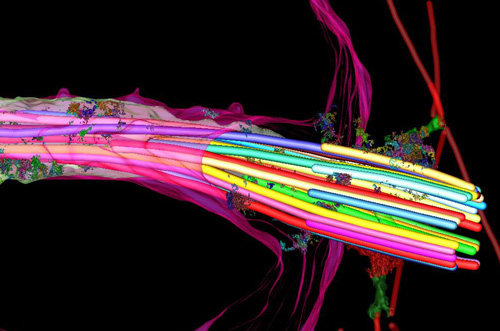
Primary cilia is formed by centrioles project from the surface of most vertebrate cells, and is involved in the response to specific developmental signals, including Hedgehog (Hh) ligands. Bangs et al. demonstrate the time and location that primary cilia appear in the mouse embryo. The article was published on Nature Cell Biology, recently.
Researchers established a transgenic mouse embryo cell line that express ARL13B fused to the red fluorescent protein mCherry in cilia, and Centrin 2 fused to GFP in centrosomes. At E6.0, primary cilia first appear in the epiblast on cells, following by the present on all derivatives of the epiblast. On the other hand, extraembryonic cell lines, such as visceral endoderm and trophectoderm lineages, are absent of cilia but have centrosomes. The results match with what they found in vivo. In addition, extraembryonic endoderm (XEN) stem cells, which lack cilia, can form cilia after inhibiting cilium disassembly pathway, AURKA-HDAC6. The absence of cilia on XEN stem cells indicates these cells are unable to respond to Hh ligands, which triggered the Hh signaling pathway to mediate embryo development. The findings suggest the ability of cells response to Hh ligands is regulated by the distribution of primary cilia in the placenta and yolk sac, therefore, influences embryo development.
Reference:
Nat Cell Biol. 2015 Jan 19. doi: 10.1038/ncb3091.
Related Products
| Cat.No. | Product Name | Information |
|---|---|---|
| S1454 | PHA-680632 | PHA-680632 is a potent inhibitor of Aurora A, Aurora B and Aurora C with IC50 of 27 nM, 135 nM and 120 nM, respectively. It has 10- to 200-fold higher IC50 for FGFR1, FLT3, LCK, PLK1, STLK2, and VEGFR2/3. |
| S1133 | Alisertib (MLN8237) | Alisertib (MLN8237) is a selective Aurora A inhibitor with IC50 of 1.2 nM in a cell-free assay. It has >200-fold higher selectivity for Aurora A than Aurora B. Alisertib induces cell cycle arrest, apoptosis and autophagy. Phase 3. |
| S1109 | BI 2536 | BI-2536 is a potent Plk1 inhibitor with IC50 of 0.83 nM in a cell-free assay. BI-2536 inhibits Bromodomain 4 (BRD4) with Kd of 37 nM and potently suppresses c-Myc expression. BI-2536 induces apoptosis and attenuates autophagy. Phase 2. |
Related Targets
Products are for research use only. Not for human use. We do not sell to patients.
©Copyright 2013 Selleck Chemicals. All Rights Reserved.