- Bioactive Compounds
- By Signaling Pathways
- PI3K/Akt/mTOR
- Epigenetics
- Methylation
- Immunology & Inflammation
- Protein Tyrosine Kinase
- Angiogenesis
- Apoptosis
- Autophagy
- ER stress & UPR
- JAK/STAT
- MAPK
- Cytoskeletal Signaling
- Cell Cycle
- TGF-beta/Smad
- DNA Damage/DNA Repair
- Compound Libraries
- Popular Compound Libraries
- Customize Library
- Clinical and FDA-approved Related
- Bioactive Compound Libraries
- Inhibitor Related
- Natural Product Related
- Metabolism Related
- Cell Death Related
- By Signaling Pathway
- By Disease
- Anti-infection and Antiviral Related
- Neuronal and Immunology Related
- Fragment and Covalent Related
- FDA-approved Drug Library
- FDA-approved & Passed Phase I Drug Library
- Preclinical/Clinical Compound Library
- Bioactive Compound Library-I
- Bioactive Compound Library-Ⅱ
- Kinase Inhibitor Library
- Express-Pick Library
- Natural Product Library
- Human Endogenous Metabolite Compound Library
- Alkaloid Compound LibraryNew
- Angiogenesis Related compound Library
- Anti-Aging Compound Library
- Anti-alzheimer Disease Compound Library
- Antibiotics compound Library
- Anti-cancer Compound Library
- Anti-cancer Compound Library-Ⅱ
- Anti-cancer Metabolism Compound Library
- Anti-Cardiovascular Disease Compound Library
- Anti-diabetic Compound Library
- Anti-infection Compound Library
- Antioxidant Compound Library
- Anti-parasitic Compound Library
- Antiviral Compound Library
- Apoptosis Compound Library
- Autophagy Compound Library
- Calcium Channel Blocker LibraryNew
- Cambridge Cancer Compound Library
- Carbohydrate Metabolism Compound LibraryNew
- Cell Cycle compound library
- CNS-Penetrant Compound Library
- Covalent Inhibitor Library
- Cytokine Inhibitor LibraryNew
- Cytoskeletal Signaling Pathway Compound Library
- DNA Damage/DNA Repair compound Library
- Drug-like Compound Library
- Endoplasmic Reticulum Stress Compound Library
- Epigenetics Compound Library
- Exosome Secretion Related Compound LibraryNew
- FDA-approved Anticancer Drug LibraryNew
- Ferroptosis Compound Library
- Flavonoid Compound Library
- Fragment Library
- Glutamine Metabolism Compound Library
- Glycolysis Compound Library
- GPCR Compound Library
- Gut Microbial Metabolite Library
- HIF-1 Signaling Pathway Compound Library
- Highly Selective Inhibitor Library
- Histone modification compound library
- HTS Library for Drug Discovery
- Human Hormone Related Compound LibraryNew
- Human Transcription Factor Compound LibraryNew
- Immunology/Inflammation Compound Library
- Inhibitor Library
- Ion Channel Ligand Library
- JAK/STAT compound library
- Lipid Metabolism Compound LibraryNew
- Macrocyclic Compound Library
- MAPK Inhibitor Library
- Medicine Food Homology Compound Library
- Metabolism Compound Library
- Methylation Compound Library
- Mouse Metabolite Compound LibraryNew
- Natural Organic Compound Library
- Neuronal Signaling Compound Library
- NF-κB Signaling Compound Library
- Nucleoside Analogue Library
- Obesity Compound Library
- Oxidative Stress Compound LibraryNew
- Plant Extract Library
- Phenotypic Screening Library
- PI3K/Akt Inhibitor Library
- Protease Inhibitor Library
- Protein-protein Interaction Inhibitor Library
- Pyroptosis Compound Library
- Small Molecule Immuno-Oncology Compound Library
- Mitochondria-Targeted Compound LibraryNew
- Stem Cell Differentiation Compound LibraryNew
- Stem Cell Signaling Compound Library
- Natural Phenol Compound LibraryNew
- Natural Terpenoid Compound LibraryNew
- TGF-beta/Smad compound library
- Traditional Chinese Medicine Library
- Tyrosine Kinase Inhibitor Library
- Ubiquitination Compound Library
-
Cherry Picking
You can personalize your library with chemicals from within Selleck's inventory. Build the right library for your research endeavors by choosing from compounds in all of our available libraries.
Please contact us at [email protected] to customize your library.
You could select:
- Antibodies
- Bioreagents
- qPCR
- 2x SYBR Green qPCR Master Mix
- 2x SYBR Green qPCR Master Mix(Low ROX)
- 2x SYBR Green qPCR Master Mix(High ROX)
- Protein Assay
- Protein A/G Magnetic Beads for IP
- Anti-Flag magnetic beads
- Anti-Flag Affinity Gel
- Anti-Myc magnetic beads
- Anti-HA magnetic beads
- Magnetic Separator
- Poly DYKDDDDK Tag Peptide lyophilized powder
- Protease Inhibitor Cocktail
- Protease Inhibitor Cocktail (EDTA-Free, 100X in DMSO)
- Phosphatase Inhibitor Cocktail (2 Tubes, 100X)
- Cell Biology
- Cell Counting Kit-8 (CCK-8)
- Animal Experiment
- Mouse Direct PCR Kit (For Genotyping)
- New Products
- Contact Us
PHA-793887
PHA-793887 is a novel and potent inhibitor of CDK2, CDK5 and CDK7 with IC50 of 8 nM, 5 nM and 10 nM. It is greater than 6-fold more selective for CDK2, 5, and 7 than CDK1, 4, and 9. PHA-793887 induces cell-cycle arrest and apoptosis. Phase 1.

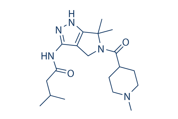
PHA-793887 Chemical Structure
CAS No. 718630-59-2
Purity & Quality Control
Batch:
S148701
DMSO]72 mg/mL]false]Ethanol]72 mg/mL]false]Water]Insoluble]false
Purity:
99.64%
99.64
PHA-793887 Related Products
Signaling Pathway
Cell Data
| Cell Lines | Assay Type | Concentration | Incubation Time | Formulation | Activity Description | PMID |
|---|---|---|---|---|---|---|
| human A2780 cells | Proliferation assay | 72 h | Antiproliferative activity against human A2780 cells after 72 hrs by fluorescence assay, IC50=0.09 μM | 20153204 | ||
| human HCT116 cells | Proliferation assay | 72 h | Antiproliferative activity against human HCT116 cells after 72 hrs by SRB assay, IC50=0.163 μM | 20153204 | ||
| human COLO205 cells | Proliferation assay | 72 h | Antiproliferative activity against human COLO205 cells after 72 hrs by SRB assay, IC50=0.188 μM | 20153204 | ||
| human C-433 cells | Proliferation assay | 72 h | Antiproliferative activity against human C-433 cells after 72 hrs by SRB assay, IC50=0.285 μM | 20153204 | ||
| human DU145 cells | Proliferation assay | 72 h | Antiproliferative activity against human DU145 cells after 72 hrs by SRB assay, IC50=0.303 μM | 20153204 | ||
| human A375 cells | Proliferation assay | 72 h | Antiproliferative activity against human A375 cells after 72 hrs by SRB assay, IC50=0.396 μM | 20153204 | ||
| human PC3 cells | Proliferation assay | 72 h | Antiproliferative activity against human PC3 cells after 72 hrs by SRB assay, IC50=0.601 μM | 20153204 | ||
| human MCF7 cells | Proliferation assay | 72 h | Antiproliferative activity against human MCF7 cells after 72 hrs by SRB assay, IC50=1.284 μM | 20153204 | ||
| human BxPC3 cells | Proliferation assay | 72 h | Antiproliferative activity against human BxPC3 cells after 72 hrs by SRB assay, IC50=3.444 μM | 20153204 | ||
| human A2780 cells | Function assay | 3 μM | Inhibition of CDK in human A2780 cells assessed as accumulation of hypophosphorylated form of retinoblastoma protein at 3 uM by immunohistochemistry | 20153204 | ||
| Click to View More Cell Line Experimental Data | ||||||
Biological Activity
| Description | PHA-793887 is a novel and potent inhibitor of CDK2, CDK5 and CDK7 with IC50 of 8 nM, 5 nM and 10 nM. It is greater than 6-fold more selective for CDK2, 5, and 7 than CDK1, 4, and 9. PHA-793887 induces cell-cycle arrest and apoptosis. Phase 1. | |||||||||||
|---|---|---|---|---|---|---|---|---|---|---|---|---|
| Features | Multi-CDK inhibitor. | |||||||||||
| Targets |
|
| In vitro | ||||
| In vitro | PHA-793887 has low activity against CDK1, CDK4, CDK9 and GSK3β with IC50 of 60 nM, 62 nM, 138 nM and 79 nM, respectively. PHA-793887 inhibits cell proliferation of many tumor cell lines, including A2780, HCT-116, COLO-205, C-433, DU-145, A375, PC3, MCF-7, and BX-PC3, with IC50 of 88 nM–3.4 μM. PHA-793887 (1 μM) shows a decrease in the S phase, a subsequent increase of the G1 phase and a slight accumulation of G2/M phase in A2780 cells. PHA-793887 (3 μM) significantly increases G2/M phase and reduces DNA synthsis. [1] PHA-793887 is cytotoxic for leukemic cell lines, including K562, KU812, KCL22, and TOM1, with IC50 of 0.3–7 μM, but it is not cytotoxic for normal unstimulated peripheral blood mononuclear cells or CD34+ hematopoietic stem cells. In colony assays, PHA-793887 shows very high activity against leukemia cell lines with IC50 less than 0.1 μM. PHA-793887 induces cell-cycle arrest, inhibits Rb and nucleophosmin phosphorylation, and modulates cyclin E and cdc6 expression at 0.2−1 μM and induces apoptosis at 5 μM. [2] | |||
|---|---|---|---|---|
| Kinase Assay | CDK Kinase Assay | |||
| The biochemical activity of compounds is determined by incubation with specific enzymes and substrates, followed by quantitation of the phosphorylated product. PHA-793887 (1.5 nM–10 μM) is incubated for 30−90 min at room temperature in the presence of ATP/33P-γ-ATP mix, substrate, and the specific enzyme (0.7−100 nM) in a final volume of 30 μL of kinase buffer, using 96 U bottom plates. After incubation, the reaction is stopped and the phosphorylated substrate is separated from nonincorporated radioactive ATP using SPA beads, Dowex resin, or Multiscreen phosphocellulose filter as follows: (1) For SPA Assays. The reaction is stopped by the addition of 100 μL of PBS + 32 mM EDTA + 0.1% Triton X-100 + 500 μM ATP, containing 1 mg of streptavidin-coated SPA beads. After 20 min of incubation for substrate capture, 100 μL of the reaction mixture is transferred into Optiplate 96-well plates containing 100 μL of 5 M CsCl, left to stand for 4 hours to allow stratification of beads to the top of the plate, and counted using TopCount to measure substrate-incorporated phosphate. (2) For Dowex Resin Assay. An amount of 150 μL of resin/formate, pH 3.00, is added to stop the reaction and capture unreacted 33P-γ-ATP, separating it from the phosphorylated substrate in solution. After 60 min of rest, 50 μL of supernatant is transferred to Optiplate 96-well plates. After the additon of 150 μL of Microscint 40, the radioactivity is counted in the TopCount. (3) For Multiscreen Assay. The reaction is stopped with the addition of 10 μL of EDTA (150 mM). An amount of 100 μL is transferred to a MultiScreen plate to allow substrate binding to phosphocellulose filter. Plates are then washed three times with 100 μL of H2PO4 (75 mM) filtered by a MultiScreen filtration system, and dried. After the additon of 100 μL of Microscint 0, radioactivity is counted in the TopCount. IC50 values are obtained by nonlinear regression analysis. | ||||
| Cell Research | Cell lines | A2780 cells | ||
| Concentrations | 0.1 nM-1 μM, dissolved in DMSO | |||
| Incubation Time | 72 hours | |||
| Method | Cells are seeded into 96- or 384-wells plates at final concentration ranging from 1 × 10⁴ to 3 × 10⁴ per cm2. After 24 hours, cells are treated using serial dilution of PHA-793887. At 72 hours after the treatment, the amount of cells are evaluated using the CellTiter-Glo assay. IC50 values are calculated using a sygmoidal fitt | |||
| In Vivo | ||
| In vivo | PHA-793887 (10–30 mg/kg) shows good efficacy in the human ovarian A2780, colon HCT-116, and pancreatic BX-PC3 carcinoma xenograft models. [1] PHA-793887 (20 mg/kg) is effective in xenograft models of K562 and HL60 cells, primary leukemic disseminated model, and a high-burden disseminated ALL-2 model derived from a relapsed Philadelphia-positive acute lymphoid leukemia patient. [2] | |
|---|---|---|
| Animal Research | Animal Models | Mouse xenograft models of human ovarian A2780, colon HCT-116 and pancreatic BX-PC3 carcinoma |
| Dosages | 10, 20, and 30 mg/kg | |
| Administration | Intravenous injection once daily | |
Chemical Information & Solubility
| Molecular Weight | 361.48 | Formula | C19H31N5O2 |
| CAS No. | 718630-59-2 | SDF | Download PHA-793887 SDF |
| Smiles | CC(C)CC(=O)NC1=NNC2=C1CN(C2(C)C)C(=O)C3CCN(CC3)C | ||
| Storage (From the date of receipt) | |||
|
In vitro |
DMSO : 72 mg/mL ( (199.18 mM) Moisture-absorbing DMSO reduces solubility. Please use fresh DMSO.) Ethanol : 72 mg/mL Water : Insoluble |
Molecular Weight Calculator |
|
In vivo Add solvents to the product individually and in order. |
In vivo Formulation Calculator |
||||
Preparing Stock Solutions
Molarity Calculator
In vivo Formulation Calculator (Clear solution)
Step 1: Enter information below (Recommended: An additional animal making an allowance for loss during the experiment)
mg/kg
g
μL
Step 2: Enter the in vivo formulation (This is only the calculator, not formulation. Please contact us first if there is no in vivo formulation at the solubility Section.)
% DMSO
%
% Tween 80
% ddH2O
%DMSO
%
Calculation results:
Working concentration: mg/ml;
Method for preparing DMSO master liquid: mg drug pre-dissolved in μL DMSO ( Master liquid concentration mg/mL, Please contact us first if the concentration exceeds the DMSO solubility of the batch of drug. )
Method for preparing in vivo formulation: Take μL DMSO master liquid, next addμL PEG300, mix and clarify, next addμL Tween 80, mix and clarify, next add μL ddH2O, mix and clarify.
Method for preparing in vivo formulation: Take μL DMSO master liquid, next add μL Corn oil, mix and clarify.
Note: 1. Please make sure the liquid is clear before adding the next solvent.
2. Be sure to add the solvent(s) in order. You must ensure that the solution obtained, in the previous addition, is a clear solution before proceeding to add the next solvent. Physical methods such
as vortex, ultrasound or hot water bath can be used to aid dissolving.
Tech Support
Answers to questions you may have can be found in the inhibitor handling instructions. Topics include how to prepare stock solutions, how to store inhibitors, and issues that need special attention for cell-based assays and animal experiments.
Tel: +1-832-582-8158 Ext:3
If you have any other enquiries, please leave a message.
* Indicates a Required Field
Tags: buy PHA-793887 | PHA-793887 supplier | purchase PHA-793887 | PHA-793887 cost | PHA-793887 manufacturer | order PHA-793887 | PHA-793887 distributor


