- Bioactive Compounds
- By Signaling Pathways
- PI3K/Akt/mTOR
- Epigenetics
- Methylation
- Immunology & Inflammation
- Protein Tyrosine Kinase
- Angiogenesis
- Apoptosis
- Autophagy
- ER stress & UPR
- JAK/STAT
- MAPK
- Cytoskeletal Signaling
- Cell Cycle
- TGF-beta/Smad
- DNA Damage/DNA Repair
- Compound Libraries
- Popular Compound Libraries
- Customize Library
- Clinical and FDA-approved Related
- Bioactive Compound Libraries
- Inhibitor Related
- Natural Product Related
- Metabolism Related
- Cell Death Related
- By Signaling Pathway
- By Disease
- Anti-infection and Antiviral Related
- Neuronal and Immunology Related
- Fragment and Covalent Related
- FDA-approved Drug Library
- FDA-approved & Passed Phase I Drug Library
- Preclinical/Clinical Compound Library
- Bioactive Compound Library-I
- Bioactive Compound Library-Ⅱ
- Kinase Inhibitor Library
- Express-Pick Library
- Natural Product Library
- Human Endogenous Metabolite Compound Library
- Alkaloid Compound LibraryNew
- Angiogenesis Related compound Library
- Anti-Aging Compound Library
- Anti-alzheimer Disease Compound Library
- Antibiotics compound Library
- Anti-cancer Compound Library
- Anti-cancer Compound Library-Ⅱ
- Anti-cancer Metabolism Compound Library
- Anti-Cardiovascular Disease Compound Library
- Anti-diabetic Compound Library
- Anti-infection Compound Library
- Antioxidant Compound Library
- Anti-parasitic Compound Library
- Antiviral Compound Library
- Apoptosis Compound Library
- Autophagy Compound Library
- Calcium Channel Blocker LibraryNew
- Cambridge Cancer Compound Library
- Carbohydrate Metabolism Compound LibraryNew
- Cell Cycle compound library
- CNS-Penetrant Compound Library
- Covalent Inhibitor Library
- Cytokine Inhibitor LibraryNew
- Cytoskeletal Signaling Pathway Compound Library
- DNA Damage/DNA Repair compound Library
- Drug-like Compound Library
- Endoplasmic Reticulum Stress Compound Library
- Epigenetics Compound Library
- Exosome Secretion Related Compound LibraryNew
- FDA-approved Anticancer Drug LibraryNew
- Ferroptosis Compound Library
- Flavonoid Compound Library
- Fragment Library
- Glutamine Metabolism Compound Library
- Glycolysis Compound Library
- GPCR Compound Library
- Gut Microbial Metabolite Library
- HIF-1 Signaling Pathway Compound Library
- Highly Selective Inhibitor Library
- Histone modification compound library
- HTS Library for Drug Discovery
- Human Hormone Related Compound LibraryNew
- Human Transcription Factor Compound LibraryNew
- Immunology/Inflammation Compound Library
- Inhibitor Library
- Ion Channel Ligand Library
- JAK/STAT compound library
- Lipid Metabolism Compound LibraryNew
- Macrocyclic Compound Library
- MAPK Inhibitor Library
- Medicine Food Homology Compound Library
- Metabolism Compound Library
- Methylation Compound Library
- Mouse Metabolite Compound LibraryNew
- Natural Organic Compound Library
- Neuronal Signaling Compound Library
- NF-κB Signaling Compound Library
- Nucleoside Analogue Library
- Obesity Compound Library
- Oxidative Stress Compound LibraryNew
- Plant Extract Library
- Phenotypic Screening Library
- PI3K/Akt Inhibitor Library
- Protease Inhibitor Library
- Protein-protein Interaction Inhibitor Library
- Pyroptosis Compound Library
- Small Molecule Immuno-Oncology Compound Library
- Mitochondria-Targeted Compound LibraryNew
- Stem Cell Differentiation Compound LibraryNew
- Stem Cell Signaling Compound Library
- Natural Phenol Compound LibraryNew
- Natural Terpenoid Compound LibraryNew
- TGF-beta/Smad compound library
- Traditional Chinese Medicine Library
- Tyrosine Kinase Inhibitor Library
- Ubiquitination Compound Library
-
Cherry Picking
You can personalize your library with chemicals from within Selleck's inventory. Build the right library for your research endeavors by choosing from compounds in all of our available libraries.
Please contact us at [email protected] to customize your library.
You could select:
- Antibodies
- Bioreagents
- qPCR
- 2x SYBR Green qPCR Master Mix
- 2x SYBR Green qPCR Master Mix(Low ROX)
- 2x SYBR Green qPCR Master Mix(High ROX)
- Protein Assay
- Protein A/G Magnetic Beads for IP
- Anti-Flag magnetic beads
- Anti-Flag Affinity Gel
- Anti-Myc magnetic beads
- Anti-HA magnetic beads
- Magnetic Separator
- Poly DYKDDDDK Tag Peptide lyophilized powder
- Protease Inhibitor Cocktail
- Protease Inhibitor Cocktail (EDTA-Free, 100X in DMSO)
- Phosphatase Inhibitor Cocktail (2 Tubes, 100X)
- Cell Biology
- Cell Counting Kit-8 (CCK-8)
- Animal Experiment
- Mouse Direct PCR Kit (For Genotyping)
- New Products
- Contact Us
Apoptosis Activator 2
Apoptosis Activator 2 strongly induces caspase-3 activation, PARP cleavage, and DNA fragmentation which leads to the destruction of cells (Apaf-1 dependent) with IC50 of ~4 μM, inactive to HMEC, PREC, or MCF-10A cells.

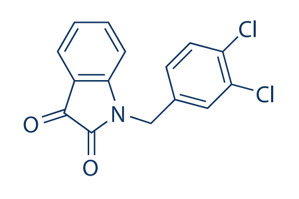
Apoptosis Activator 2 Chemical Structure
CAS No. 79183-19-0
Purity & Quality Control
Apoptosis Activator 2 Related Products
| Related Targets | Caspase-1 Caspase-3 Caspase-8 Caspase-9 Caspase-4 Capase-7 Caspase-2 Caspase-5 Caspase-6 Caspase-10 Caspase-7 | Click to Expand |
|---|---|---|
| Related Products | Z-VAD-FMK Belnacasan (VX-765) Z-DEVD-FMK Q-VD-Oph Z-IETD-FMK Emricasan (IDN-6556) Ac-DEVD-CHO Z-LEHD-FMK TFA Z-VAD(OH)-FMK (Caspase Inhibitor VI) PAC-1 Z-YVAD-FMK Tasisulam Phenoxodiol (Haginin E) | Click to Expand |
| Related Compound Libraries | Autophagy Compound Library Apoptosis Compound Library Ferroptosis Compound Library Pyroptosis Compound Library Mitochondria-Targeted Compound Library | Click to Expand |
Signaling Pathway
Biological Activity
| Description | Apoptosis Activator 2 strongly induces caspase-3 activation, PARP cleavage, and DNA fragmentation which leads to the destruction of cells (Apaf-1 dependent) with IC50 of ~4 μM, inactive to HMEC, PREC, or MCF-10A cells. | |
|---|---|---|
| Targets |
|
| In vitro | ||||
| In vitro | Apoptosis Activator 2 (20 μM) at the reduced cyto c concentration increases the fraction of Apaf-1 in the apoptosome by 1.5-fold to 33%. Apoptosis Activator 2 increases the extent of caspase-3 activation at the reduced level of cyto c and caspase-3 activation by 4-fold. Apoptosis Activator 2 strongly indues caspase-3 activation, PARP cleavage, and DNA fragmentation, and finally killing cells with an IC50 of 4 μM. Apoptosis Activator 2 induces apoptosis of PBL, HUVEC, Jurkat, Molt-4, CCRF-CEM, BT-549, MDA-MB-468 and NCI-H23 with of IC50 of 50 μM, 43 μM, 4 μM, 6 μM, 9 μM, 20 μM, 44 μM and 35 μM. Apoptosis Activator 2 exerts a cytostatic effect on the majority of tumor cell lines tested, inhibiting cell growth by 50-100% at 10 μM in 40 of 48 cell lines tested. [1] Apoptosis Activator 2 induces cell death by triggering apoptosome formation. The level of En1 expression does not have a significant influence on the survival rates of Ventral midbrain cultures for Apoptosis Activator 2 (-8.1 ± 6.0%). Survival rate is not significantly altered if the other three reagents are employed (-10.7 ± 4.7%) for Apoptosis Activator 2. [2] Apoptosis activator 2 (10 μM) induces apoptosis in AGS cells as evaluated by apoptotic DNA ladder and Tunel assay. Apoptosis activator 2 (10 μM) enhances the induction of apoptosis by anti TROP2 conjugated liposomes. [3] Cyclohexamide (10 μg/mL) or zVAD (50 μM) significantly protects against Apoptosis Activator 2 toxicity in neuroneal cultures. Apoptosis activator 2 (3 μM) results in numerous neurones with pyknotic nuclei suggestive of cell death involving apoptosis. DHT (10 nM) or E2 (10 nM) significantly protects against Apoptosis Activator 2 toxicity in neuroneal cultures. [4] | |||
|---|---|---|---|---|
| Kinase Assay | Cell-Free Apoptosis Assay | |||
| HeLa cell cytoplasmic extracts are prepared according to previously published reports. Apoptosis Activator 2 in DMSO are distributed into 96-well microtiter plates at a final concentration of 1 mM (final DMSO concentration is 1% vol/vol). To each well is added 250 μg of total protein from cytoplasmic extracts in HEB buffer (50 mM Hepes, pH 7.4/50 mM KCl/5 mM EGTA/2 mM MgCl), with 2 mM DTT, 2 μM cyto c, and 0.5 μM DEVD-AFC (Asp-Glu-Val-Asp-7-amino-4-trifluoromethylcoumarin) substrate in a total of 150 μL. Plates are incubated at 37 ℃, and fluorescence is read in a LJL Biosystems plate reader at 10-min intervals. | ||||
| Cell Research | Cell lines | Neurones | ||
| Concentrations | ~3 μM | |||
| Incubation Time | 2 hours | |||
| Method | All viable cells within the defined field of a microscope reticle grid are counted using a manual mechanical counter by an experimenter blinded to condition. Cells are scored viable on the basis of both positive staining with the vital dye calcein acetoxymethyl ester and the morphological criterion of a smooth, spherical soma. Counts of viable cells are made in four non-overlapping fields per culture well with each condition represented by 3 separate wells. The number of viable cells counts per well for vehicle-treated control conditions ranged from 100-200. All experiments are repeated in at least 3 independent culture preparations. Raw cell count data are statistically analyzed with one-way ANOVA, followed by between group comparisons using the Fisher LSD test (significance indicated by P < 0.05). Cell viability is presented graphically as a percentage of live cells in the vehicle-treated control condition. | |||
Chemical Information & Solubility
| Molecular Weight | 306.14 | Formula | C15H9Cl2NO2 |
| CAS No. | 79183-19-0 | SDF | Download Apoptosis Activator 2 SDF |
| Smiles | C1=CC=C2C(=C1)C(=O)C(=O)N2CC3=CC(=C(C=C3)Cl)Cl | ||
| Storage (From the date of receipt) | |||
|
In vitro |
DMSO : 61 mg/mL ( (199.25 mM) Moisture-absorbing DMSO reduces solubility. Please use fresh DMSO.) Water : Insoluble Ethanol : Insoluble |
Molecular Weight Calculator |
|
In vivo Add solvents to the product individually and in order. |
In vivo Formulation Calculator |
||||
Preparing Stock Solutions
Molarity Calculator
In vivo Formulation Calculator (Clear solution)
Step 1: Enter information below (Recommended: An additional animal making an allowance for loss during the experiment)
mg/kg
g
μL
Step 2: Enter the in vivo formulation (This is only the calculator, not formulation. Please contact us first if there is no in vivo formulation at the solubility Section.)
% DMSO
%
% Tween 80
% ddH2O
%DMSO
%
Calculation results:
Working concentration: mg/ml;
Method for preparing DMSO master liquid: mg drug pre-dissolved in μL DMSO ( Master liquid concentration mg/mL, Please contact us first if the concentration exceeds the DMSO solubility of the batch of drug. )
Method for preparing in vivo formulation: Take μL DMSO master liquid, next addμL PEG300, mix and clarify, next addμL Tween 80, mix and clarify, next add μL ddH2O, mix and clarify.
Method for preparing in vivo formulation: Take μL DMSO master liquid, next add μL Corn oil, mix and clarify.
Note: 1. Please make sure the liquid is clear before adding the next solvent.
2. Be sure to add the solvent(s) in order. You must ensure that the solution obtained, in the previous addition, is a clear solution before proceeding to add the next solvent. Physical methods such
as vortex, ultrasound or hot water bath can be used to aid dissolving.
Tech Support
Answers to questions you may have can be found in the inhibitor handling instructions. Topics include how to prepare stock solutions, how to store inhibitors, and issues that need special attention for cell-based assays and animal experiments.
Tel: +1-832-582-8158 Ext:3
If you have any other enquiries, please leave a message.
* Indicates a Required Field
Tags: buy Apoptosis Activator 2 | Apoptosis Activator 2 supplier | purchase Apoptosis Activator 2 | Apoptosis Activator 2 cost | Apoptosis Activator 2 manufacturer | order Apoptosis Activator 2 | Apoptosis Activator 2 distributor


