- Bioactive Compounds
- By Signaling Pathways
- PI3K/Akt/mTOR
- Epigenetics
- Methylation
- Immunology & Inflammation
- Protein Tyrosine Kinase
- Angiogenesis
- Apoptosis
- Autophagy
- ER stress & UPR
- JAK/STAT
- MAPK
- Cytoskeletal Signaling
- Cell Cycle
- TGF-beta/Smad
- DNA Damage/DNA Repair
- Compound Libraries
- Popular Compound Libraries
- Customize Library
- Clinical and FDA-approved Related
- Bioactive Compound Libraries
- Inhibitor Related
- Natural Product Related
- Metabolism Related
- Cell Death Related
- By Signaling Pathway
- By Disease
- Anti-infection and Antiviral Related
- Neuronal and Immunology Related
- Fragment and Covalent Related
- FDA-approved Drug Library
- FDA-approved & Passed Phase I Drug Library
- Preclinical/Clinical Compound Library
- Bioactive Compound Library-I
- Bioactive Compound Library-Ⅱ
- Kinase Inhibitor Library
- Express-Pick Library
- Natural Product Library
- Human Endogenous Metabolite Compound Library
- Alkaloid Compound LibraryNew
- Angiogenesis Related compound Library
- Anti-Aging Compound Library
- Anti-alzheimer Disease Compound Library
- Antibiotics compound Library
- Anti-cancer Compound Library
- Anti-cancer Compound Library-Ⅱ
- Anti-cancer Metabolism Compound Library
- Anti-Cardiovascular Disease Compound Library
- Anti-diabetic Compound Library
- Anti-infection Compound Library
- Antioxidant Compound Library
- Anti-parasitic Compound Library
- Antiviral Compound Library
- Apoptosis Compound Library
- Autophagy Compound Library
- Calcium Channel Blocker LibraryNew
- Cambridge Cancer Compound Library
- Carbohydrate Metabolism Compound LibraryNew
- Cell Cycle compound library
- CNS-Penetrant Compound Library
- Covalent Inhibitor Library
- Cytokine Inhibitor LibraryNew
- Cytoskeletal Signaling Pathway Compound Library
- DNA Damage/DNA Repair compound Library
- Drug-like Compound Library
- Endoplasmic Reticulum Stress Compound Library
- Epigenetics Compound Library
- Exosome Secretion Related Compound LibraryNew
- FDA-approved Anticancer Drug LibraryNew
- Ferroptosis Compound Library
- Flavonoid Compound Library
- Fragment Library
- Glutamine Metabolism Compound Library
- Glycolysis Compound Library
- GPCR Compound Library
- Gut Microbial Metabolite Library
- HIF-1 Signaling Pathway Compound Library
- Highly Selective Inhibitor Library
- Histone modification compound library
- HTS Library for Drug Discovery
- Human Hormone Related Compound LibraryNew
- Human Transcription Factor Compound LibraryNew
- Immunology/Inflammation Compound Library
- Inhibitor Library
- Ion Channel Ligand Library
- JAK/STAT compound library
- Lipid Metabolism Compound LibraryNew
- Macrocyclic Compound Library
- MAPK Inhibitor Library
- Medicine Food Homology Compound Library
- Metabolism Compound Library
- Methylation Compound Library
- Mouse Metabolite Compound LibraryNew
- Natural Organic Compound Library
- Neuronal Signaling Compound Library
- NF-κB Signaling Compound Library
- Nucleoside Analogue Library
- Obesity Compound Library
- Oxidative Stress Compound LibraryNew
- Plant Extract Library
- Phenotypic Screening Library
- PI3K/Akt Inhibitor Library
- Protease Inhibitor Library
- Protein-protein Interaction Inhibitor Library
- Pyroptosis Compound Library
- Small Molecule Immuno-Oncology Compound Library
- Mitochondria-Targeted Compound LibraryNew
- Stem Cell Differentiation Compound LibraryNew
- Stem Cell Signaling Compound Library
- Natural Phenol Compound LibraryNew
- Natural Terpenoid Compound LibraryNew
- TGF-beta/Smad compound library
- Traditional Chinese Medicine Library
- Tyrosine Kinase Inhibitor Library
- Ubiquitination Compound Library
-
Cherry Picking
You can personalize your library with chemicals from within Selleck's inventory. Build the right library for your research endeavors by choosing from compounds in all of our available libraries.
Please contact us at info@selleckchem.com to customize your library.
You could select:
- Antibodies
- Bioreagents
- qPCR
- 2x SYBR Green qPCR Master Mix
- 2x SYBR Green qPCR Master Mix(Low ROX)
- 2x SYBR Green qPCR Master Mix(High ROX)
- Protein Assay
- Protein A/G Magnetic Beads for IP
- Anti-Flag magnetic beads
- Anti-Flag Affinity Gel
- Anti-Myc magnetic beads
- Anti-HA magnetic beads
- Poly DYKDDDDK Tag Peptide lyophilized powder
- Protease Inhibitor Cocktail
- Protease Inhibitor Cocktail (EDTA-Free, 100X in DMSO)
- Phosphatase Inhibitor Cocktail (2 Tubes, 100X)
- Cell Biology
- Cell Counting Kit-8 (CCK-8)
- Animal Experiment
- Mouse Direct PCR Kit (For Genotyping)
- New Products
- Contact Us
Perifosine
Synonyms: KRX-0401, NSC639966, D21266
Home PI3K/Akt/mTOR Akt inhibitor Perifosine
For research use only.
Perifosine is a novel Akt inhibitor with IC50 of 4.7 μM in MM.1S cells, targets pleckstrin homology domain of Akt. Phase 3.

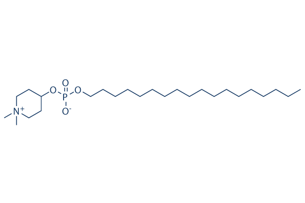
Perifosine Chemical Structure
CAS No. 157716-52-4
Purity & Quality Control
Batch:
Purity: >97%
97
Perifosine Related Products
| Related Targets | Akt1 Akt2 Akt3 | Click to Expand |
|---|---|---|
| Related Products | MK-2206 2HCl SC79 Capivasertib (AZD5363) GSK690693 Ipatasertib (GDC-0068) Triciribine (API-2) CCT128930 Afuresertib (GSK2110183) A-674563 HCl AT7867 Oridonin Akti-1/2 PHT-427 AT13148 Miransertib (ARQ 092) HCl Uprosertib (GSK2141795) Miransertib (ARQ-092) SC66 Deguelin ML-9 HCl | Click to Expand |
| Related Compound Libraries | Kinase Inhibitor Library PI3K/Akt Inhibitor Library Apoptosis Compound Library Cell Cycle compound library NF-κB Signaling Compound Library | Click to Expand |
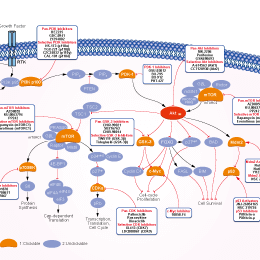
Signaling Pathway
Cell Data
| Cell Lines | Assay Type | Concentration | Incubation Time | Formulation | Activity Description | PMID |
|---|---|---|---|---|---|---|
| HL-60 | Apoptosis Asssay | 10 μM | 24/48 h | induces apoptosis time-dependently | 20130960 | |
| MOLM | Apoptosis Asssay | 10 μM | 24/48 h | induces apoptosis time-dependently | 20130960 | |
| OCI | Apoptosis Asssay | 10 μM | 24/48 h | induces apoptosis time-dependently | 20130960 | |
| BJAB | Apoptosis Asssay | 10 μM | 24/48 h | induces apoptosis time-dependently | 20130960 | |
| MAVER | Apoptosis Asssay | 10 μM | 24/48 h | induces apoptosis time-dependently | 20130960 | |
| SKW6.4 | Apoptosis Asssay | 10 μM | 24/48 h | induces apoptosis time-dependently | 20130960 | |
| HL-60 | Growth Inhibition Assay | 2-10 μM | 48 h | inhibits cell growth in a dose dependent manner | 20130960 | |
| MOLM | Growth Inhibition Assay | 2-10 μM | 48 h | inhibits cell growth in a dose dependent manner | 20130960 | |
| OCI | Growth Inhibition Assay | 2-10 μM | 48 h | inhibits cell growth in a dose dependent manner | 20130960 | |
| BJAB | Growth Inhibition Assay | 2-10 μM | 48 h | inhibits cell growth in a dose dependent manner | 20130960 | |
| MAVER | Growth Inhibition Assay | 2-10 μM | 48 h | inhibits cell growth in a dose dependent manner | 20130960 | |
| SKW6.4 | Growth Inhibition Assay | 2-10 μM | 48 h | inhibits cell growth in a dose dependent manner | 20130960 | |
| A2780cis | Growth Inhibition Assay | 0-20 μM | 48/72 h | IC50 = 6 μm | 20405296 | |
| A2780 | Growth Inhibition Assay | 0-20 μM | 48/72 h | IC50 = 3 μm | 20405296 | |
| SKOV3 | Growth Inhibition Assay | 0-40 μM | 72 h | IC50~30 μM, inhibits cell growth in a dose dependent manner | 20405296 | |
| PA-1 | Growth Inhibition Assay | 0-40 μM | 72 h | IC50~25 μM, inhibits cell growth in a dose dependent manner | 20405296 | |
| OAW-42 | Growth Inhibition Assay | 0-40 μM | 72 h | IC50~10 μM, inhibits cell growth in a dose dependent manner | 20405296 | |
| Bel-7402 | Apoptosis Asssay | 5/10/20 μM | 24/48 h | induces apoptosis at the long-time exposure | 20842425 | |
| HepG2 | Apoptosis Asssay | 5/10/20 μM | 24/48 h | induces apoptosis at the long-time exposure | 20842425 | |
| Bel-7402 | Function Assay | 5/10/20 μM | 24 h | results in the accumulation of cell number in the G2/M phase | 20842425 | |
| HepG2 | Function Assay | 5/10/20 μM | 24 h | results in the accumulation of cell number in the G2/M phase | 20842425 | |
| Bel-7402 | Growth Inhibition Assay | 5/10/20/40 μM | 24/48/72 h | inhibits cell growth in both time and dose dependent manner | 20842425 | |
| HepG2 | Growth Inhibition Assay | 5/10/20/40 μM | 24/48/72 h | inhibits cell growth in both time and dose dependent manner | 20842425 | |
| CWR22RV1 | Function Assay | 5 μM | 24 h | reduced phosphorylation of Akt significantly | 21496273 | |
| CWR22RV1 | Apoptosis Asssay | 10 μM | 24 h | enhances radiation induced apoptosis | 21496273 | |
| CWR22RV1 | Cell Viability Assay | 10 μM | 24 h | increases sensitivity of human CWR22RV1 cells to radiation | 21496273 | |
| A498 | Growth Inhibition Assay | 0-20 μM | 72 h | inhibits cell growth in a dose dependent manner | 21644050 | |
| 769-P | Growth Inhibition Assay | 0-20 μM | 72 h | inhibits cell growth in a dose dependent manner | 21644050 | |
| CAKI-1 | Growth Inhibition Assay | 0-20 μM | 72 h | inhibits cell growth in a dose dependent manner | 21644050 | |
| 786-O | Growth Inhibition Assay | 0-20 μM | 72 h | inhibits cell growth in a dose dependent manner | 21644050 | |
| 786-0 | Growth Inhibition Assay | 0-40 μM | 72 h | IC50~5 μM | 21644050 | |
| 769-P | Growth Inhibition Assay | 0-40 μM | 72 h | IC50~5-10 μM | 21644050 | |
| CAKI-1 | Growth Inhibition Assay | 0-40 μM | 72 h | IC50~10 μM | 21644050 | |
| A498 | Growth Inhibition Assay | 0-40 μM | 72 h | inhibits cell growth in a dose dependent manner | 21644050 | |
| HT-29 | Cytotoxicity Assay | 5 μM | 48 h | enhances paclitaxel induced ovarian cancer cell death | 21775054 | |
| A2780 | Cytotoxicity Assay | 5 μM | 48 h | enhances paclitaxel induced ovarian cancer cell death | 21775054 | |
| SKOV3 | Cytotoxicity Assay | 5 μM | 48 h | enhances paclitaxel induced ovarian cancer cell death | 21775054 | |
| CaOV3 | Cell Viability Assay | 1/5/10 μM | 48 h | decreases cell viability in a dose dependent manner cotreated with paclitaxel | 21775054 | |
| OCUT1 | Function Assay | 3 μm | 24 h | causes a dramatic increase in G2/M phase | 22090271 | |
| K1 | Growth Inhibition Assay | 0.1-3 μM | 5 d | inhibits cell growth in a dose dependent manner | 22090271 | |
| OCUT1 | Growth Inhibition Assay | 0.1-3 μM | 5 d | inhibits cell growth in a dose dependent manner | 22090271 | |
| K562 | Function Assay | 20 μM | 48 h | induces autophagy | 22407228 | |
| HL-60 | Function Assay | 2.5/5/10 μM | 24 h | induces the phosphorylation of JNK1/2 in a dose dependent manner | 22407228 | |
| Kasumi-1 | Function Assay | 2.5/5/10 μM | 24 h | induces the phosphorylation of JNK1/2 in a dose dependent manner | 22407228 | |
| HL-60 | Function Assay | 2.5/5/10 μM | 24 h | decreases Akt and p-Akt levels dose-dependently | 22407228 | |
| Kasumi-1 | Function Assay | 2.5/5/10 μM | 24 h | decreases Akt and p-Akt levels dose-dependently | 22407228 | |
| HL-60 | Apoptosis Asssay | 10 μM | 24 h | induces apoptosis | 22407228 | |
| Kasumi-1 | Apoptosis Asssay | 10 μM | 24 h | induces apoptosis | 22407228 | |
| HL-60 | Cell Viability Assay | 0-20 μM | 24/48 h | decreases cell viability in both dose and time dependent manner | 22407228 | |
| Kasumi-1 | Cell Viability Assay | 0-20 μM | 24/48 h | decreases cell viability in both dose and time dependent manner | 22407228 | |
| BON1 | Function Assay | 7.5/10 μM | 8 h | decreases the expression of the anti-apoptotic proteins BCL2 and Bcl-XL | 22499437 | |
| BON1 | Apoptosis Asssay | 0-10 μM | 24 h | induces apoptosis dose dependently | 22499437 | |
| BON1 | Cell Viability Assay | 0-100 μM | 24/72 h | decreases cell viability in both dose and time dependent manner | 22499437 | |
| GOT1 | Cell Viability Assay | 0-100 μM | 24/72 h | decreases cell viability in both dose and time dependent manner | 22499437 | |
| NCI-H727 | Cell Viability Assay | 0-100 μM | 24/72 h | decreases cell viability in both dose and time dependent manner | 22499437 | |
| MCAS | Growth Inhibition Assay | IC50=12.5 μM | 23877012 | |||
| A2780S | Growth Inhibition Assay | IC50=14.5 μM | 23877012 | |||
| OVCAR5 | Growth Inhibition Assay | IC50=6.7 μM | 23877012 | |||
| A2780CP | Growth Inhibition Assay | IC50=7.6 μM | 23877012 | |||
| HeyA8 | Growth Inhibition Assay | IC50=24.3 μM | 23877012 | |||
| OVCAR8 | Growth Inhibition Assay | IC50=31.1 μM | 23877012 | |||
| M41R | Growth Inhibition Assay | IC50=19.8 μM | 23877012 | |||
| M41 | Growth Inhibition Assay | IC50=24.7 μM | 23877012 | |||
| TykNuR | Growth Inhibition Assay | IC50=5.5 μM | 23877012 | |||
| TykNu | Growth Inhibition Assay | IC50=3.5 μM | 23877012 | |||
| MGC803 | Function Assay | 0.75/10 μM | 48 h | decreases p-Akt (Ser 473), p-GSK3β (Ser 9), and C-MYC levels | 23912246 | |
| SGC7901 | Function Assay | 0.75/10 μM | 48 h | decreases p-Akt (Ser 473), p-GSK3β (Ser 9), and C-MYC levels | 23912246 | |
| U87MG | Cell Viability Assay | 0-25 μM | 24-96 h | decreases cell viability in both dose and time dependent manner | 24065522 | |
| AsPC-1 | Function Assay | 0.5 μM | 24 h | inhibits Akt, S6K1, and Erk1/2 phosphorylation | 24519751 | |
| MIA | Function Assay | 0.5 μM | 24 h | inhibits Akt, S6K1, and Erk1/2 phosphorylation | 24519751 | |
| PANC-1 | Function Assay | 0.5 μM | 24 h | inhibits Akt, S6K1, and Erk1/2 phosphorylation | 24519751 | |
| AsPC-1 | Growth Inhibition Assay | 0-25 μM | 72 h | inhibits cell growth in a dose dependent manner | 24519751 | |
| MIA | Growth Inhibition Assay | 0-25 μM | 72 h | inhibits cell growth in a dose dependent manner | 24519751 | |
| PANC-1 | Growth Inhibition Assay | 0-25 μM | 72 h | inhibits cell growth in a dose dependent manner | 24519751 | |
| Ema | Growth Inhibition Assay | 0.1–100 μM | 48 h | IC50=58.7 μM | 24881508 | |
| UL-1 | Growth Inhibition Assay | 0.1–100 μM | 48 h | IC50=7.01 μM | 24881508 | |
| CLBL-1 | Growth Inhibition Assay | 0.1–100 μM | 48 h | IC50=33.0 μM | 24881508 | |
| GL-1 | Growth Inhibition Assay | 0.1–100 μM | 48 h | IC50=9.91 μM | 24881508 | |
| MDA-MB-231 | Growth Inhibition Assay | 0-10 μM | 48 h | EC50=1.13 ± 0.07 μM | 25293576 | |
| HCC1806 | Growth Inhibition Assay | 0-10 μM | 48 h | EC50=2.84 ± 0.07 μM | 25293576 | |
| RMG2 | Apoptosis Asssay | 30 μM | 24 h | induces apoptosis | 25519148 | |
| RMG1 | Apoptosis Asssay | 30 μM | 24 h | induces apoptosis | 25519148 | |
| A2780 | Cell Viability Assay | 1-30 μM | 48 h | decreases cell viability in a dose dependent manner | 25519148 | |
| SKOV3 | Cell Viability Assay | 1-30 μM | 48 h | decreases cell viability in a dose dependent manner | 25519148 | |
| OVISE | Cell Viability Assay | 1-30 μM | 48 h | decreases cell viability in a dose dependent manner | 25519148 | |
| RMG2 | Cell Viability Assay | 1-30 μM | 48 h | decreases cell viability in a dose dependent manner | 25519148 | |
| HAC2 | Cell Viability Assay | 1-30 μM | 72 h | decreases cell viability in a dose dependent manner | 25519148 | |
| KOC7C | Cell Viability Assay | 1-30 μM | 72 h | decreases cell viability in a dose dependent manner | 25519148 | |
| RMG2 | Cell Viability Assay | 1-30 μM | 72 h | decreases cell viability in a dose dependent manner | 25519148 | |
| RMG1 | Cell Viability Assay | 1-30 μM | 72 h | decreases cell viability in a dose dependent manner | 25519148 | |
| H460 | Function Assay | 3 μM | 8 h | blocks mTORC1, and ERK-MAPK activation combined with MEK-162 | 25697899 | |
| A549 | Function Assay | 3 μM | 8 h | blocks mTORC1, and ERK-MAPK activation combined with MEK-162 | 25697899 | |
| H460 | Function Assay | 3 μM | 8 h | blocks AKT activation | 25697899 | |
| A549 | Function Assay | 3 μM | 8 h | blocks AKT activation | 25697899 | |
| H460 | Apoptosis Asssay | 1/3 μM | 48 h | induces apoptosis | 25697899 | |
| A549 | Apoptosis Asssay | 1/3 μM | 48 h | induces apoptosis | 25697899 | |
| H460 | Growth Inhibition Assay | 0.3-10 μM | 24/72 h | inhibits cell growth in both time and dose dependent manner | 25697899 | |
| A549 | Growth Inhibition Assay | 0.3-10 μM | 24/72 h | inhibits cell growth in both time and dose dependent manner | 25697899 | |
| U-87 MG | Growth Inhibition Assay | 20/40 μM | 24/48 h | inhibits cell growth in both time and dose dependent manner | 25934232 | |
| HepG2 | Growth Inhibition Assay | 20/40 μM | 24/48 h | inhibits cell growth in both time and dose dependent manner | 25934232 | |
| U-87 MG | Function Assay | 20 μM | 6/24 h | increases the autophagic flux at 6 h while inhibits this flux at 24h | 25934232 | |
| HepG2 | Function Assay | 20 μM | 6/24 h | decreases LC3-II degradation from 6 h | 25934232 | |
| U-87 MG | Function Assay | 20 μM | 6/24 h | increases the levels of LC3-II cotreated with CQ | 25934232 | |
| HepG2 | Function Assay | 20 μM | 6/24 h | increases the levels of LC3-II cotreated with CQ | 25934232 | |
| U-87 MG | Function Assay | 20 μM | 24 h | increases double-membrane bound structures | 25934232 | |
| HepG2 | Function Assay | 20 μM | 24 h | produces an intense cytoplasmic vacuolization corresponding to a notable dilatation of the ER cisterns | 25934232 | |
| T24 BC | Apoptosis Asssay | 2.5 μM | 24 h | sensitizes BC cells to sorafenib-induced apoptotic | 26097873 | |
| T24 BC | Cell Viability Assay | 0.5/1/2.5 μM | 24 h | enhances sorafenib-induced cell viability decrease | 26097873 | |
| T24 BC | Function Assay | 0.5/1/2.5 μM | 3 h | reduces the basal CB tyrosine phosphorylation levels in a dose-dependent manner | 26097873 | |
| RBL2H3 | Function assay | Toxicity in rat RBL2H3 cells, MTD=25μM | 20153565 | |||
| PC3 | Growth inhibition assay | Growth inhibition of human PC3 cells by sulforhodamine B assay, GI50=0.44μM | 21543141 | |||
| NUGC3 | Growth inhibition assay | Growth inhibition of human NUGC3 cells by sulforhodamine B assay, GI50=0.54μM | 21543141 | |||
| HCT15 | Growth inhibition assay | Growth inhibition of human HCT15 cells by sulforhodamine B assay, GI50=1.25μM | 21543141 | |||
| MDA-MB-231 | Growth inhibition assay | Growth inhibition of human MDA-MB-231 cells by sulforhodamine B assay, GI50=2.86μM | 21543141 | |||
| NCI-H23 | Growth inhibition assay | Growth inhibition of human NCI-H23 cells by sulforhodamine B assay, GI50=4.21μM | 21543141 | |||
| ACHN | Growth inhibition assay | Growth inhibition of human ACHN cells by sulforhodamine B assay, GI50=4.56μM | 21543141 | |||
| A549 | Function assay | 30 mins | Inhibition of Akt phosphorylation in insulin-stimulated human A549 cells treated 2 hrs before insulin stimulation measured after 30 mins by ELISA, IC50=5.3μM | 22138309 | ||
| A549 | Cytotoxicity assay | 24 hrs | Cytotoxicity against human A549 cells after 24 hrs by FACS analysis, IC50=7μM | 22138309 | ||
| KATO III | Cytotoxicity assay | 24 hrs | Cytotoxicity against human KATO III cells after 24 hrs by FACS analysis, IC50=12.8μM | 22138309 | ||
| MCF7 | Cytotoxicity assay | 24 hrs | Cytotoxicity against human MCF7 cells after 24 hrs by FACS analysis, IC50=13.3μM | 22138309 | ||
| PC3 | Growth inhibition assay | Growth inhibition of human PC3 cells by SRB assay, GI50=0.44μM | 23266181 | |||
| NUGC3 | Growth inhibition assay | Growth inhibition of human NUGC3 cells by SRB assay, GI50=0.54μM | 23266181 | |||
| HCT15 | Growth inhibition assay | Growth inhibition of human HCT15 cells by SRB assay, GI50=1.25μM | 23266181 | |||
| MDA-MB-231 | Growth inhibition assay | Growth inhibition of human MDA-MB-231 cells by SRB assay, GI50=2.86μM | 23266181 | |||
| NCI-H23 | Growth inhibition assay | Growth inhibition of human NCI-H23 cells by SRB assay, GI50=4.21μM | 23266181 | |||
| ACHN | Growth inhibition assay | Growth inhibition of human ACHN cells by SRB assay, GI50=4.56μM | 23266181 | |||
| A549 | Function assay | 2 hrs | Inhibition of Akt phosphorylation in human insulin-stimulated A549 cells incubated for 2 hrs prior to insulin-induction measured after 30 mins by ELISA, IC50=5.3μM | 23415083 | ||
| A549 | Cytotoxicity assay | Cytotoxicity against human A549 cells by flow cytometric analysis, IC50=7μM | 23415083 | |||
| KATO III | Cytotoxicity assay | Cytotoxicity against human KATO III cells by flow cytometric analysis, IC50=12.8μM | 23415083 | |||
| MCF7 | Cytotoxicity assay | Cytotoxicity against human MCF7 cells by flow cytometric analysis, IC50=13.3μM | 23415083 | |||
| PC3 | Antiproliferative assay | Antiproliferative activity against human PC3 cells by SRB assay, GI50=0.44μM | 23567950 | |||
| NUGC3 | Antiproliferative assay | Antiproliferative activity against human NUGC3 cells by SRB assay, GI50=0.54μM | 23567950 | |||
| HCT15 | Antiproliferative assay | Antiproliferative activity against human HCT15 cells by SRB assay, GI50=1.25μM | 23567950 | |||
| MDA-MB-231 | Antiproliferative assay | Antiproliferative activity against human MDA-MB-231 cells by SRB assay, GI50=2.86μM | 23567950 | |||
| NCI-H23 | Antiproliferative assay | Antiproliferative activity against human NCI-H23 cells by SRB assay, GI50=4.21μM | 23567950 | |||
| ACHN | Antiproliferative assay | Antiproliferative activity against human ACHN cells by SRB assay, GI50=4.56μM | 23567950 | |||
| PC3 | Growth inhibition assay | 48 hrs | Growth inhibition of human PC3 cells after 48 hrs by SRB assay, GI50=0.44μM | 24095759 | ||
| NUGC3 | Growth inhibition assay | 48 hrs | Growth inhibition of human NUGC3 cells after 48 hrs by SRB assay, GI50=0.54μM | 24095759 | ||
| HCT15 | Growth inhibition assay | 48 hrs | Growth inhibition of human HCT15 cells after 48 hrs by SRB assay, GI50=1.25μM | 24095759 | ||
| MDA-MB-231 | Growth inhibition assay | 48 hrs | Growth inhibition of human MDA-MB-231 cells after 48 hrs by SRB assay, GI50=2.86μM | 24095759 | ||
| NCI-H23 | Growth inhibition assay | 48 hrs | Growth inhibition of human NCI-H23 cells after 48 hrs by SRB assay, GI50=4.21μM | 24095759 | ||
| ACHN | Growth inhibition assay | 48 hrs | Growth inhibition of human ACHN cells after 48 hrs by SRB assay, GI50=4.56μM | 24095759 | ||
| A549 | Cytotoxicity assay | 24 to 72 hrs | Cytotoxicity against human A549 cells after 24 to 72 hrs by haemocytometry, IC50=4.17μM | 24900620 | ||
| Rosetta cells | Function assay | Inhibition of wild-type human P38alpha MAPK expressed in Escherichia coli Rosetta cells, IC50=1.2μM | 31274316 | |||
| Click to View More Cell Line Experimental Data | ||||||
Biological Activity
| Description | Perifosine is a novel Akt inhibitor with IC50 of 4.7 μM in MM.1S cells, targets pleckstrin homology domain of Akt. Phase 3. | ||
|---|---|---|---|
| Targets |
|
| In vitro | ||||
| In vitro | Perifosine develops anti-proliferative properties with IC50 of 0.6-8.9 μM in immortalized keratinocytes (HaCaT), and head and neck squamous carcinoma cells. [1] Perifosine strongly reduces phosphorylation levels of Akt and extracellular signal-regulated kinase (Erk) 1/2, induces cell cycle arrest in G1 and G2, and causes dose-dependent growth inhibition of mouse glial progenitors. [2] Perifosine (10 μM) completely inhibits the phosphorylation of Akt in MM.1S cells. [3] A recent study demonstrates Perifosine induces cell cycle arrest and apoptosis in human hepatocellular carcinoma cell lines by blockade of Akt phosphorylation. [4] |
|||
|---|---|---|---|---|
| Kinase Assay | Akt kinase assay | |||
| MM.1S cells are cultured in the presence or absence of perifosine (5 μM, 6 hours) and then stimulated with IL-6 (20 ng/mL, 10 minutes). In vitro akt kinase assay is then carried out using the Akt Kinase Assay Kit. | ||||
| Cell Research | Cell lines | Human glioma cell lines | ||
| Concentrations | 0, 15, 30 and 45 μM | |||
| Incubation Time | 48 hours | |||
| Method | Cells are incubated in the medium with 10% FCS for 48 hours with indicated concentration of Periosine. Cell viability is determined by the MTT assay. (Cell Proliferation Kit I; Roche). The absorbance at 590 nm is recorded using the 96-well plate reader. |
|||
| Experimental Result Images | Methods | Biomarkers | Images | PMID |
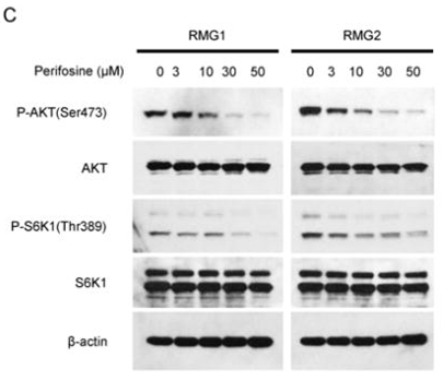
| Western blot | p-AKT / AKT / p-S6K1 / S6K1 PARP p-mTOR / mTOR / Raptor / Rictor / p-p70S6K / p70S6K / p-4EBP1 / 4EBP1 / c-Myc / Cyclin D1 p-PDK1 / p-GSK3α/β / p-S6R |

|
25519148 | |
| Growth inhibition assay | Cell viability |

|
28332584 | |
| In Vivo | ||
| In vivo | Perifosine in combination reduces tumor proliferation (a PDGF-driven gliomagenesis) in vivo. The results indicate that Perifosine is an effective drug in gliomas in which Akt and Ras-Erk 1/2 pathways are frequently activated, and may be new candidate for glima treatment in the clinic. [2] Both oral daily and weekly administration of Perifosine significantly reduce human MM tumor growth and increase survival, compared with control animals treated with PBS vehicle only. [3] Perifosine induces thrombocytosis and leukocytosis and increases myelopoiesis in murine marrow and spleen, whereas it causes apoptosis in myeloma xenografts. [5] |
|
|---|---|---|
| Animal Research | Animal Models | MM.1S MM cells are inoculated subcutaneously in the right flank of Beige-nude-xid (BNX) mice (5 to 6 weeks old). |
| Dosages | 250 mg/kg/wk or 36 mg/kg/d | |
| Administration | Oral gavage | |
| NCT Number | Recruitment | Conditions | Sponsor/Collaborators | Start Date | Phases |
|---|---|---|---|---|---|
| NCT01224730 | Completed | Cancer |
AEterna Zentaris |
January 24 2012 | Phase 1 |
| NCT01049841 | Completed | Pediatric Solid Tumors |
Memorial Sloan Kettering Cancer Center|University of Wisconsin Madison|Duke University|NATL COMP CA NETWORK|Pfizer|AEterna Zentaris |
January 2010 | Phase 1 |
| NCT01048580 | Completed | Colon Cancer |
AEterna Zentaris|SCRI Development Innovations LLC |
October 2009 | Phase 1 |
| NCT00776867 | Completed | Solid Tumors |
Memorial Sloan Kettering Cancer Center|University of Wisconsin Madison|Duke University|AEterna Zentaris |
October 2008 | Phase 1 |
|
Chemical Information & Solubility
| Molecular Weight | 461.66 | Formula | C25H52NO4P |
| CAS No. | 157716-52-4 | SDF | Download Perifosine SDF |
| Smiles | CCCCCCCCCCCCCCCCCCOP(=O)([O-])OC1CC[N+](CC1)(C)C | ||
| Storage (From the date of receipt) | |||
|
In vitro |
Water : 92 mg/mL Ethanol : 92 mg/mL DMSO : Insoluble ( Moisture-absorbing DMSO reduces solubility. Please use fresh DMSO.) |
Molecular Weight Calculator |
|
In vivo Add solvents to the product individually and in order. |
In vivo Formulation Calculator |
|||||
Preparing Stock Solutions
Molarity Calculator
In vivo Formulation Calculator (Clear solution)
Step 1: Enter information below (Recommended: An additional animal making an allowance for loss during the experiment)
mg/kg
g
μL
Step 2: Enter the in vivo formulation (This is only the calculator, not formulation. Please contact us first if there is no in vivo formulation at the solubility Section.)
% DMSO
%
% Tween 80
% ddH2O
%DMSO
%
Calculation results:
Working concentration: mg/ml;
Method for preparing DMSO master liquid: mg drug pre-dissolved in μL DMSO ( Master liquid concentration mg/mL, Please contact us first if the concentration exceeds the DMSO solubility of the batch of drug. )
Method for preparing in vivo formulation: Take μL DMSO master liquid, next addμL PEG300, mix and clarify, next addμL Tween 80, mix and clarify, next add μL ddH2O, mix and clarify.
Method for preparing in vivo formulation: Take μL DMSO master liquid, next add μL Corn oil, mix and clarify.
Note: 1. Please make sure the liquid is clear before adding the next solvent.
2. Be sure to add the solvent(s) in order. You must ensure that the solution obtained, in the previous addition, is a clear solution before proceeding to add the next solvent. Physical methods such
as vortex, ultrasound or hot water bath can be used to aid dissolving.
Tech Support
Answers to questions you may have can be found in the inhibitor handling instructions. Topics include how to prepare stock solutions, how to store inhibitors, and issues that need special attention for cell-based assays and animal experiments.
Tel: +1-832-582-8158 Ext:3
If you have any other enquiries, please leave a message.
* Indicates a Required Field


