- Bioactive Compounds
- By Signaling Pathways
- PI3K/Akt/mTOR
- Epigenetics
- Methylation
- Immunology & Inflammation
- Protein Tyrosine Kinase
- Angiogenesis
- Apoptosis
- Autophagy
- ER stress & UPR
- JAK/STAT
- MAPK
- Cytoskeletal Signaling
- Cell Cycle
- TGF-beta/Smad
- DNA Damage/DNA Repair
- Compound Libraries
- Popular Compound Libraries
- Customize Library
- Clinical and FDA-approved Related
- Bioactive Compound Libraries
- Inhibitor Related
- Natural Product Related
- Metabolism Related
- Cell Death Related
- By Signaling Pathway
- By Disease
- Anti-infection and Antiviral Related
- Neuronal and Immunology Related
- Fragment and Covalent Related
- FDA-approved Drug Library
- FDA-approved & Passed Phase I Drug Library
- Preclinical/Clinical Compound Library
- Bioactive Compound Library-I
- Bioactive Compound Library-Ⅱ
- Kinase Inhibitor Library
- Express-Pick Library
- Natural Product Library
- Human Endogenous Metabolite Compound Library
- Alkaloid Compound LibraryNew
- Angiogenesis Related compound Library
- Anti-Aging Compound Library
- Anti-alzheimer Disease Compound Library
- Antibiotics compound Library
- Anti-cancer Compound Library
- Anti-cancer Compound Library-Ⅱ
- Anti-cancer Metabolism Compound Library
- Anti-Cardiovascular Disease Compound Library
- Anti-diabetic Compound Library
- Anti-infection Compound Library
- Antioxidant Compound Library
- Anti-parasitic Compound Library
- Antiviral Compound Library
- Apoptosis Compound Library
- Autophagy Compound Library
- Calcium Channel Blocker LibraryNew
- Cambridge Cancer Compound Library
- Carbohydrate Metabolism Compound LibraryNew
- Cell Cycle compound library
- CNS-Penetrant Compound Library
- Covalent Inhibitor Library
- Cytokine Inhibitor LibraryNew
- Cytoskeletal Signaling Pathway Compound Library
- DNA Damage/DNA Repair compound Library
- Drug-like Compound Library
- Endoplasmic Reticulum Stress Compound Library
- Epigenetics Compound Library
- Exosome Secretion Related Compound LibraryNew
- FDA-approved Anticancer Drug LibraryNew
- Ferroptosis Compound Library
- Flavonoid Compound Library
- Fragment Library
- Glutamine Metabolism Compound Library
- Glycolysis Compound Library
- GPCR Compound Library
- Gut Microbial Metabolite Library
- HIF-1 Signaling Pathway Compound Library
- Highly Selective Inhibitor Library
- Histone modification compound library
- HTS Library for Drug Discovery
- Human Hormone Related Compound LibraryNew
- Human Transcription Factor Compound LibraryNew
- Immunology/Inflammation Compound Library
- Inhibitor Library
- Ion Channel Ligand Library
- JAK/STAT compound library
- Lipid Metabolism Compound LibraryNew
- Macrocyclic Compound Library
- MAPK Inhibitor Library
- Medicine Food Homology Compound Library
- Metabolism Compound Library
- Methylation Compound Library
- Mouse Metabolite Compound LibraryNew
- Natural Organic Compound Library
- Neuronal Signaling Compound Library
- NF-κB Signaling Compound Library
- Nucleoside Analogue Library
- Obesity Compound Library
- Oxidative Stress Compound LibraryNew
- Plant Extract Library
- Phenotypic Screening Library
- PI3K/Akt Inhibitor Library
- Protease Inhibitor Library
- Protein-protein Interaction Inhibitor Library
- Pyroptosis Compound Library
- Small Molecule Immuno-Oncology Compound Library
- Mitochondria-Targeted Compound LibraryNew
- Stem Cell Differentiation Compound LibraryNew
- Stem Cell Signaling Compound Library
- Natural Phenol Compound LibraryNew
- Natural Terpenoid Compound LibraryNew
- TGF-beta/Smad compound library
- Traditional Chinese Medicine Library
- Tyrosine Kinase Inhibitor Library
- Ubiquitination Compound Library
-
Cherry Picking
You can personalize your library with chemicals from within Selleck's inventory. Build the right library for your research endeavors by choosing from compounds in all of our available libraries.
Please contact us at [email protected] to customize your library.
You could select:
- Antibodies
- Bioreagents
- qPCR
- 2x SYBR Green qPCR Master Mix
- 2x SYBR Green qPCR Master Mix(Low ROX)
- 2x SYBR Green qPCR Master Mix(High ROX)
- Protein Assay
- Protein A/G Magnetic Beads for IP
- Anti-Flag magnetic beads
- Anti-Flag Affinity Gel
- Anti-Myc magnetic beads
- Anti-HA magnetic beads
- Magnetic Separator
- Poly DYKDDDDK Tag Peptide lyophilized powder
- Protease Inhibitor Cocktail
- Protease Inhibitor Cocktail (EDTA-Free, 100X in DMSO)
- Phosphatase Inhibitor Cocktail (2 Tubes, 100X)
- Cell Biology
- Cell Counting Kit-8 (CCK-8)
- Animal Experiment
- Mouse Direct PCR Kit (For Genotyping)
- New Products
- Contact Us
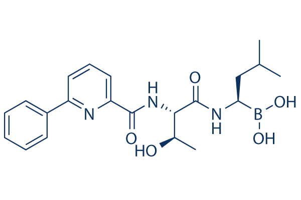
Delanzomib
Synonyms: CEP-18770
Delanzomib is an orally active inhibitor of the chymotrypsin-like activity of proteasome with IC50 of 3.8 nM, with only marginal inhibition of the tryptic and peptidylglutamyl activities of the proteosome. Phase 1/2.

Delanzomib Chemical Structure
CAS No. 847499-27-8
Purity & Quality Control
Delanzomib Related Products
| Related Targets | 20S proteasome | Click to Expand |
|---|---|---|
| Related Products | MG132 Epoxomicin (BU-4061T) Celastrol Oprozomib ONX-0914 (PR-957) VR23 Marizomib (Salinosporamide A) PI-1840 | Click to Expand |
| Related Compound Libraries | FDA-approved Drug Library Natural Product Library Bioactive Compound Library-I Protease Inhibitor Library Ubiquitination Compound Library | Click to Expand |
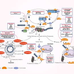
Signaling Pathway
Biological Activity
| Description | Delanzomib is an orally active inhibitor of the chymotrypsin-like activity of proteasome with IC50 of 3.8 nM, with only marginal inhibition of the tryptic and peptidylglutamyl activities of the proteosome. Phase 1/2. | ||
|---|---|---|---|
| Targets |
|
| In vitro | ||||
| In vitro | CEP-18770 demonstrates marginal prevention of the tryptic and peptidyl gultamyl activities of the protesome.[1] CEP-18770 inhibits A2780 ovarian cancer cells, PC3 prostate cancer, H460, LoVo colon cancer, RPMI8226 multiple myeloma cancer and HS-Sultan anaplastic non-Hodgkin lymphoma with IC50 values of 13.7, 22.2, 34.2 11.3, 5.6 and 8.2 nM, respectively.[1] CEP-18770 blocks the ubiquitin-proteasome pathway in several MM and in the chronic myelogenous leukemia cell line, K562. CEP-18770 causes an accumulation of ubiquitinated proteins over 4 to 8 hours.[1] IκBα degradation is completely blocked by pretreatment with CEP-18770. CEP-18770 significantly inhibits high levels of NF-κB activity in both RPMI-8226 and U266 cells. The time- and concentration-dependent suppression of NF-kB DNA-binding activity in MM cell lines by CEP-18770 leads to a decrease of expression of several NF-κB-modulated genes mediating the growth and survival of tumor cells including IkBα itself, the X-chromosome-linked inhibitor-of-apoptosis protein (XIAP), the pro-inflammatory cytokines TNF-α and interleukin-1β (IL-1β), the intracellular adhesion molecule (ICAM1), and the pro-angiogeneic factor vascular endothelial growth factor. [1] The expression of these NF-κB–mediated genes are associated with more favorable clinical responsiveness to this agent, highlighting their potential prognostic value in response to CEP-18770 exposure. [1] The proapoptotic activity of CEP-18770 against MM is not limited solely to tumor-derived MM cell lines, but extends to primary MM explants from relapsed or refractory patients. [1] In addition, CEP-18770 in combination produces synergistic inhibition of MM cell viability in vitro.[2] |
|||
|---|---|---|---|---|
| Kinase Assay | Probing proteasome activity in cell extracts | |||
| Human multiple myeloma cells are washed twice with cold phosphate-buffered saline, pelleted and lysed with one volume of glass beads (<106 microns, acid-washed) and an equal volume of homogenization buffer (50 mM Tris (pH 7.4), 1 mM dithiothreitol, 5 mM MgCl2, 2 mM ATP and 250 mM sucrose) by vortexing at high speed for 15-30 min at 4 °C. Beads, membrane fractions, nuclei and cell debris are then removed from the supernatant by centrifugation at 16,000g for 5 min. The protein content of extracts is quantitated using the Bradford assay. Proteasome activity is assayed as described below. Equal amounts (typically 60 g) of protein are denatured by boiling in reducing sample buffer, separated by 12.5% SDS-PAGE and electrotransferred onto polyvinylidene difluoride (PVDF) membranes. Immunoblotting is performed using a dansyl-sulfonamidohexanoyl polyclonal antibody (1:7,500, rabbit) and horseradish peroxidase–coupled goat or swine anti-rabbit secondary antibody followed by enhanced chemiluminescence. | ||||
| Cell Research | Cell lines | HMEC and TEC cells | ||
| Concentrations | 0-100 nM | |||
| Incubation Time | 6 hours | |||
| Method | HMEC and TEC cells are seeded into 24-well plates at a density of 104 cells/well in DMEM supplemented with 5% FCS. After incubation with proteasome inhibitors (48 hours), cells are washed, air dried, and stained with crystal violet as described. Cell number is determined, in duplicate samples, on the basis of a standard curve obtained with known cell numbers. All experiments are performed in triplicate. In vitro formation of capillary-like structures is studied on cells (4 × 10⁴ cells/well in DMEM supplemented with 5% FCS. After incubation with proteasome inhibitors (48 hours), cells are washed (cells/well in 24-well plates) and seeded onto Matrigel-coated wells in DMEM containing 0.25% BSA. HMEC and TEC cells (5 × 10³ per well), suspended in 200 μL DMEM with 5% FCS (positive control), serum-free medium (negative control), are layered onto the Matrigel surface in the presence or absence of proteasome inhibitor CEP-18770. Cells are observed with a microscope and experimental results are then recorded after a 6-hour incubation at 37 °C. Data is analyzed, as the mean (× 1 SD) of total length of capillary-like structures, by the Micro-Image system and is expressed as mm/field by the computer analysis system in 5 different fields at 100 × magnification in duplicated wells for 4 different experiments. |
|||
| In Vivo | ||
| In vivo | CEP-18770 reveals sustained dose-related relative tumor weight inhibition. CEP-18770 leads to dose-related complete tumor regressions, which results in a 50% incidence of CR at its maximally tolerated dose (MTD) of 1.2 mg/kg intravenously. [1] CEP-18770 reveals dose-related increase in the incidence of tumor-free mice by the completion of these studies (120 days after tumor transplantation). Oral administration of CEP-18770 produces a marked decrease in tumor weight and notable dose-related incidence of complete tumor regression with minimal changes in animal body weight over the course of the 120 day studies. [1]Equiactive doses of CEP-18770 reveal a greater and more sustained dose-related inhibition of tumor proteasome activity, corresponding temporarily with maximum induction of caspase-3 and 7 activity.[1] The maximum apoptotic signal is 2.5 fold greater for CEP-18770. In contrast, proteasome inhibition profiles of CEP-18870 are comparable in the normal peripheral mouse tissues examined (liver, lungs, whole blood, and brain [no activity]) in both their magnitude and their duration.[1] No proteasome inhibition is detected in brain tissue at any time point for either CEP-18770 or. [1] Single agent CEP-18770 PO also shows marked anti-MM effects in these xenograft models[1] |
|
|---|---|---|
| Animal Research | Animal Models | Human MM RPMI 8226 subcutaneous xenograft model in SCID mice |
| Dosages | From 1.5 to 4 mg/kg, twice for 7 days to 4 weeks. | |
| Administration | Intravenously | |
| NCT Number | Recruitment | Conditions | Sponsor/Collaborators | Start Date | Phases |
|---|---|---|---|---|---|
| NCT01348919 | Completed | Multiple Myeloma |
Teva Branded Pharmaceutical Products R&D Inc. |
August 3 2011 | Phase 1|Phase 2 |
| NCT01023880 | Terminated | Multiple Myeloma |
Cephalon|Teva Branded Pharmaceutical Products R&D Inc. |
January 2010 | Phase 1|Phase 2 |
| NCT00572637 | Completed | Solid Tumors|Lymphoma Non-Hodgkin |
Ethical Oncology Science |
November 2007 | Phase 1 |
Chemical Information & Solubility
| Molecular Weight | 413.28 | Formula | C21H28BN3O5 |
| CAS No. | 847499-27-8 | SDF | Download Delanzomib SDF |
| Smiles | B(C(CC(C)C)NC(=O)C(C(C)O)NC(=O)C1=CC=CC(=N1)C2=CC=CC=C2)(O)O | ||
| Storage (From the date of receipt) | |||
|
In vitro |
DMSO : 83 mg/mL ( (200.83 mM) Moisture-absorbing DMSO reduces solubility. Please use fresh DMSO.) Ethanol : 83 mg/mL Water : Insoluble |
Molecular Weight Calculator |
|
In vivo Add solvents to the product individually and in order. |
In vivo Formulation Calculator |
||||
Preparing Stock Solutions
Molarity Calculator
In vivo Formulation Calculator (Clear solution)
Step 1: Enter information below (Recommended: An additional animal making an allowance for loss during the experiment)
mg/kg
g
μL
Step 2: Enter the in vivo formulation (This is only the calculator, not formulation. Please contact us first if there is no in vivo formulation at the solubility Section.)
% DMSO
%
% Tween 80
% ddH2O
%DMSO
%
Calculation results:
Working concentration: mg/ml;
Method for preparing DMSO master liquid: mg drug pre-dissolved in μL DMSO ( Master liquid concentration mg/mL, Please contact us first if the concentration exceeds the DMSO solubility of the batch of drug. )
Method for preparing in vivo formulation: Take μL DMSO master liquid, next addμL PEG300, mix and clarify, next addμL Tween 80, mix and clarify, next add μL ddH2O, mix and clarify.
Method for preparing in vivo formulation: Take μL DMSO master liquid, next add μL Corn oil, mix and clarify.
Note: 1. Please make sure the liquid is clear before adding the next solvent.
2. Be sure to add the solvent(s) in order. You must ensure that the solution obtained, in the previous addition, is a clear solution before proceeding to add the next solvent. Physical methods such
as vortex, ultrasound or hot water bath can be used to aid dissolving.
Tech Support
Answers to questions you may have can be found in the inhibitor handling instructions. Topics include how to prepare stock solutions, how to store inhibitors, and issues that need special attention for cell-based assays and animal experiments.
Tel: +1-832-582-8158 Ext:3
If you have any other enquiries, please leave a message.
* Indicates a Required Field
Tags: buy Delanzomib | Delanzomib supplier | purchase Delanzomib | Delanzomib cost | Delanzomib manufacturer | order Delanzomib | Delanzomib distributor


