- Bioactive Compounds
- By Signaling Pathways
- PI3K/Akt/mTOR
- Epigenetics
- Methylation
- Immunology & Inflammation
- Protein Tyrosine Kinase
- Angiogenesis
- Apoptosis
- Autophagy
- ER stress & UPR
- JAK/STAT
- MAPK
- Cytoskeletal Signaling
- Cell Cycle
- TGF-beta/Smad
- DNA Damage/DNA Repair
- Compound Libraries
- Popular Compound Libraries
- Customize Library
- Clinical and FDA-approved Related
- Bioactive Compound Libraries
- Inhibitor Related
- Natural Product Related
- Metabolism Related
- Cell Death Related
- By Signaling Pathway
- By Disease
- Anti-infection and Antiviral Related
- Neuronal and Immunology Related
- Fragment and Covalent Related
- FDA-approved Drug Library
- FDA-approved & Passed Phase I Drug Library
- Preclinical/Clinical Compound Library
- Bioactive Compound Library-I
- Bioactive Compound Library-Ⅱ
- Kinase Inhibitor Library
- Express-Pick Library
- Natural Product Library
- Human Endogenous Metabolite Compound Library
- Alkaloid Compound LibraryNew
- Angiogenesis Related compound Library
- Anti-Aging Compound Library
- Anti-alzheimer Disease Compound Library
- Antibiotics compound Library
- Anti-cancer Compound Library
- Anti-cancer Compound Library-Ⅱ
- Anti-cancer Metabolism Compound Library
- Anti-Cardiovascular Disease Compound Library
- Anti-diabetic Compound Library
- Anti-infection Compound Library
- Antioxidant Compound Library
- Anti-parasitic Compound Library
- Antiviral Compound Library
- Apoptosis Compound Library
- Autophagy Compound Library
- Calcium Channel Blocker LibraryNew
- Cambridge Cancer Compound Library
- Carbohydrate Metabolism Compound LibraryNew
- Cell Cycle compound library
- CNS-Penetrant Compound Library
- Covalent Inhibitor Library
- Cytokine Inhibitor LibraryNew
- Cytoskeletal Signaling Pathway Compound Library
- DNA Damage/DNA Repair compound Library
- Drug-like Compound Library
- Endoplasmic Reticulum Stress Compound Library
- Epigenetics Compound Library
- Exosome Secretion Related Compound LibraryNew
- FDA-approved Anticancer Drug LibraryNew
- Ferroptosis Compound Library
- Flavonoid Compound Library
- Fragment Library
- Glutamine Metabolism Compound Library
- Glycolysis Compound Library
- GPCR Compound Library
- Gut Microbial Metabolite Library
- HIF-1 Signaling Pathway Compound Library
- Highly Selective Inhibitor Library
- Histone modification compound library
- HTS Library for Drug Discovery
- Human Hormone Related Compound LibraryNew
- Human Transcription Factor Compound LibraryNew
- Immunology/Inflammation Compound Library
- Inhibitor Library
- Ion Channel Ligand Library
- JAK/STAT compound library
- Lipid Metabolism Compound LibraryNew
- Macrocyclic Compound Library
- MAPK Inhibitor Library
- Medicine Food Homology Compound Library
- Metabolism Compound Library
- Methylation Compound Library
- Mouse Metabolite Compound LibraryNew
- Natural Organic Compound Library
- Neuronal Signaling Compound Library
- NF-κB Signaling Compound Library
- Nucleoside Analogue Library
- Obesity Compound Library
- Oxidative Stress Compound LibraryNew
- Plant Extract Library
- Phenotypic Screening Library
- PI3K/Akt Inhibitor Library
- Protease Inhibitor Library
- Protein-protein Interaction Inhibitor Library
- Pyroptosis Compound Library
- Small Molecule Immuno-Oncology Compound Library
- Mitochondria-Targeted Compound LibraryNew
- Stem Cell Differentiation Compound LibraryNew
- Stem Cell Signaling Compound Library
- Natural Phenol Compound LibraryNew
- Natural Terpenoid Compound LibraryNew
- TGF-beta/Smad compound library
- Traditional Chinese Medicine Library
- Tyrosine Kinase Inhibitor Library
- Ubiquitination Compound Library
-
Cherry Picking
You can personalize your library with chemicals from within Selleck's inventory. Build the right library for your research endeavors by choosing from compounds in all of our available libraries.
Please contact us at [email protected] to customize your library.
You could select:
- Antibodies
- Bioreagents
- qPCR
- 2x SYBR Green qPCR Master Mix
- 2x SYBR Green qPCR Master Mix(Low ROX)
- 2x SYBR Green qPCR Master Mix(High ROX)
- Protein Assay
- Protein A/G Magnetic Beads for IP
- Anti-Flag magnetic beads
- Anti-Flag Affinity Gel
- Anti-Myc magnetic beads
- Anti-HA magnetic beads
- Magnetic Separator
- Poly DYKDDDDK Tag Peptide lyophilized powder
- Protease Inhibitor Cocktail
- Protease Inhibitor Cocktail (EDTA-Free, 100X in DMSO)
- Phosphatase Inhibitor Cocktail (2 Tubes, 100X)
- Cell Biology
- Cell Counting Kit-8 (CCK-8)
- Animal Experiment
- Mouse Direct PCR Kit (For Genotyping)
- New Products
- Contact Us
Nutlin-3
Nutlin-3 is a potent and selective Mdm2 (RING finger-dependent ubiquitin protein ligase for itself and p53) antagonist with IC50 of 90 nM in a cell-free assay; stabilizes p73 in p53-deficient cells.
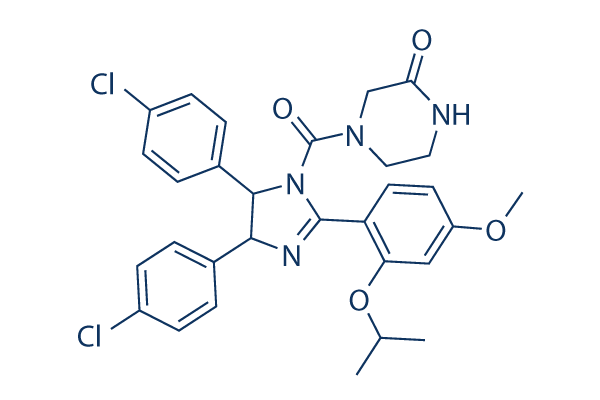
Nutlin-3 Chemical Structure
CAS No. 890090-75-2
Purity & Quality Control
Batch:
Purity:
99.59%
99.59
Products often used together with Nutlin-3
Nutlin-3 and Tozasertib combined therapy selectively kills p53-compromised cells and inhibits the proliferation of cells expressing wild-type p53.
Cheok CF, et al. Cell Death Differ. 2010 Sep;17(9):1486-500.
Nutlin-3 Related Products
| Related Targets | MDM2 MDMX p53-MDM2 interaction | Click to Expand |
|---|---|---|
| Related Products | Nutlin-3a RG-7112 Idasanutlin (RG7388) MI-773 (SAR405838) NSC 207895 NVP-CGM097 Siremadlin (HDM201) Nutlin-3b MX69 YH239-EE | Click to Expand |
| Related Compound Libraries | Autophagy Compound Library Apoptosis Compound Library Ferroptosis Compound Library Pyroptosis Compound Library Mitochondria-Targeted Compound Library | Click to Expand |
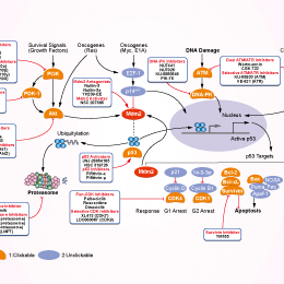
Signaling Pathway
Cell Data
| Cell Lines | Assay Type | Concentration | Incubation Time | Formulation | Activity Description | PMID |
|---|---|---|---|---|---|---|
| SMMC-7721 | Function Assay | 10 μM | 36 h | DMSO | down-regulates the protein expression level of phospho-Ser392-p53 | 24286312 |
| HuH-7 | Apoptosis Assay | 20 μM | 48 h | DMSO | induces apoptosis | 24286312 |
| SMMC-7721 | Apoptosis Assay | 20 μM | 48 h | DMSO | induces apoptosis | 24286312 |
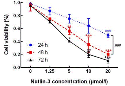
| HuH-7 | Cell Viability Assay | 1.25-20 μM | 24/48/72 h | DMSO | inhibits cell proliferation dose and time dependently | 24286312 |
| SMMC-7721 | Cell Viability Assay | 1.25-20 μM | 24/48/72 h | DMSO | inhibits cell proliferation dose and time dependently | 24286312 |
| RKO | Function Assay | 20 μM | 24 h | induces HO-1 expression at the level of transcription | 24366007 | |
| U2OS | Function Assay | 20 μM | 24 h | induces HO-1 expression at the level of transcription | 24366007 | |
| RKO | Function Assay | 20 μM | 24 h | increases the levels of the HO-1 protein as well as the p53 protein | 24366007 | |
| U2OS | Function Assay | 20 μM | 24 h | increases the levels of the HO-1 protein as well as the p53 protein | 24366007 | |
| MOLM | Function Assay | 10 μM | 24 h | upregulates the SOCS-1 expression | 24473562 | |
| OCI | Function Assay | 10 μM | 24 h | upregulates the SOCS-1 expression | 24473562 | |
| BeWo | Apoptosis Assay | 30 µM | 24 h | increases apoptosis | 24498154 | |
| BeWo | Function Assay | 30 µM | 24 h | increases p53, Mdm2, p21 and Puma at the protein level | 24498154 | |
| MOML13 | Function Assay | 10μM | 2/4 h | increases the level of p53 | 24659749 | |
| AML3 | Function Assay | 10μM | 2/4 h | increases the level of p53 | 24659749 | |
| AML2 | Function Assay | 10μM | 2/4 h | increases the level of p53 | 24659749 | |
| MOML13 | Apoptosis Assay | 2/10 μM | 24/48 h | induces apoptosis | 24659749 | |
| AML2 | Apoptosis Assay | 2/10 μM | 24/48 h | induces apoptosis | 24659749 | |
| U2OS | Function Assay | 20 μM | 24 h | increases the mRNA levels of BCL2A1, BCLXL andBCLW | 24867259 | |
| Hep3B | Apoptosis Assay | induces apoptosis | 24884809 | |||
| Huh-7 | Apoptosis Assay | induces apoptosis | 24884809 | |||
| SMMC7721 | Apoptosis Assay | induces apoptosis | 24884809 | |||
| HepG2 | Apoptosis Assay | induces apoptosis | 24884809 | |||
| Hep3B | Growth Inhibition Assay | 72 h | DMSO | IC50=20.18 ± 1.84 μM | 24884809 | |
| Huh-7 | Growth Inhibition Assay | 72 h | DMSO | IC50=33.96 ± 3.9 μM | 24884809 | |
| SMMC7721/Ac | Growth Inhibition Assay | 72 h | DMSO | IC50=55.21 ± 5.03 μM | 24884809 | |
| SMMC7721 | Growth Inhibition Assay | 72 h | DMSO | IC50=31.28 ± 4.2 μM | 24884809 | |
| HepG2/As | Growth Inhibition Assay | 72 h | DMSO | IC50=68.13 ± 9.6 μM | 24884809 | |
| HepG2 | Growth Inhibition Assay | 72 h | DMSO | IC50=35.86 ± 2.9 μM | 24884809 | |
| MOLM-13 | Function Assay | 6 μM | 6 h | DMSO | enhances the acetylation of histone H2B and heat shock proteins Hsp27 and Hsp90 | 24885082 |
| MOLM-13 | Function Assay | 6 μM | 0-8 h | DMSO | increases the levels of p53, MDM2, p21 and acetylated p53 | 24885082 |
| 115 | Function Assay | 5 μM | 48 h | DMSO | induces p53-dependent senescence | 25067787 |
| A498 | Function Assay | 5 μM | 48 h | DMSO | induces p53-dependent senescence | 25067787 |
| Caki-2 | Function Assay | 5 μM | 48 h | DMSO | induces p53-dependent senescence | 25067787 |
| ACHN | Function Assay | 5 μM | 48 h | DMSO | induces p53-dependent senescence | 25067787 |
| 115 | Growth Inhibition Assay | 5 μM | 48 h | DMSO | induces cell cycle arrest | 25067787 |
| A498 | Growth Inhibition Assay | 5 μM | 48 h | DMSO | induces cell cycle arrest | 25067787 |
| Caki-2 | Growth Inhibition Assay | 5 μM | 48 h | DMSO | induces cell cycle arrest | 25067787 |
| ACHN | Growth Inhibition Assay | 5 μM | 48 h | DMSO | induces cell cycle arrest | 25067787 |
| 115 | Function Assay | 0.5/1/5 μM | 48 h | DMSO | leads to increased expression of p53 and some p53 target genes: MDM2, and p21 | 25067787 |
| A498 | Function Assay | 0.5/1/5 μM | 48 h | DMSO | leads to increased expression of p53 and some p53 target genes: MDM2, and p21 | 25067787 |
| Caki-2 | Function Assay | 0.5/1/5 μM | 48 h | DMSO | leads to increased expression of p53 and some p53 target genes: MDM2, and p21 | 25067787 |
| ACHN | Function Assay | 0.5/1/5 μM | 48 h | DMSO | leads to increased expression of p53 and some p53 target genes: MDM2, and p21 | 25067787 |
| 117 | Cell Viability Assay | 0.5-10 μM | 0-6 d | DMSO | inhibits proliferation in a dose-dependent manner | 25067787 |
| 115 | Cell Viability Assay | 0.5-10 μM | 0-6 d | DMSO | inhibits proliferation in a dose-dependent manner | 25067787 |
| A498 | Cell Viability Assay | 0.5-10 μM | 0-6 d | DMSO | inhibits proliferation in a dose-dependent manner | 25067787 |
| Caki-2 | Cell Viability Assay | 0.5-10 μM | 0-6 d | DMSO | inhibits proliferation in a dose-dependent manner | 25067787 |
| ACHN | Cell Viability Assay | 0.5-10 μM | 0-6 d | DMSO | inhibits proliferation in a dose-dependent manner | 25067787 |
| MCF7 | Function Assay | 2.5 µM | 48 h | DMSO | decreases the homologous DSB repair frequencies | 25085902 |
| MCF7 | Cell Viability Assay | 2.5 µM | 5 d | DMSO | sensitizes MCF7 to PARP inhibition | 25085902 |
| RAW 264.7 | Function Assay | 10 μM | 30 min | inhibits LPS-induced NO production | 25172547 | |
| RAW 264.7 | Function Assay | 10 μM | 30 min | reduces the LPS-augmented the NF-κB luciferase reporter gene activity | 25172547 | |
| RAW 264.7 | Function Assay | 10 μM | 30 min | prevents the p53 reduction in response to LPS | 25172547 | |
| SUM102PT | Function Assay | 10 μM | 24 h | DMSO | inhibits TGF-β3-induced EPHB2 mRNA and protein expression | 25257729 |
| SK-BR-7 | Function Assay | 10 μM | 24 h | DMSO | inhibits TGF-β3-induced EPHB2 mRNA and protein expression | 25257729 |
| MCF-10CA1a | Function Assay | 10 μM | 24 h | DMSO | inhibits TGF-β3-induced EPHB2 mRNA and protein expression | 25257729 |
| MCF-10CA1a | Function Assay | 10 μM | 24 h | DMSO | decreases the TGF-β3-induced mRNA levels ofMMP2, MMP9, and integrin β 3 | 25257729 |
| MCF-10A1 | Function Assay | 10 μM | 24/48 h | DMSO | inhibits migration of normal breast epithelial | 25257729 |
| MCF-10CA1a | Function Assay | 10 μM | 48 h | DMSO | inhibits basal invasion and reduced TGF-β3-induced invasion to basal levels | 25257729 |
| HCT116 | Function Assay | 10 µM | 24 h | causes a p53-dependent tetraploid G1-arrest in diploid HCT116 clones D3 and D8 | 25380055 | |
| A2780 | Function Assay | 10 μM | 21h | DMSO | decreases the FoxM1 levels | 25426548 |
| NCI-H23 | Function Assay | 10 μM | 21h | DMSO | decreases the FoxM1 levels | 25426548 |
| A2780 | Function Assay | 10 μM | 21h | DMSO | increases p53 protein levels | 25426548 |
| NCI-H23 | Function Assay | 10 μM | 21h | DMSO | increases p53 protein levels | 25426548 |
| OVCAR10 | Function Assay | 10 μM | 21h | DMSO | increases p53 protein levels | 25426548 |
| MCF-7 | Function Assay | 10 μM | 0-24 h | induces p53 and p21/Cip1 | 25482373 | |
| SMMC-7721 | Function Assay | 10 μM | 36 h | DMSO | causes the ectopic expression of IFI16 | 25544361 |
| SMMC-7721 | Function Assay | 10 μM | 48 h | DMSO | increases the expression level of IFNB1 mRNA | 25544361 |
| SMMC-7721 | Function Assay | 10 μM | 48 h | DMSO | induces the chromatin-bound protein IFI16 to partially localize in the cytoplasm | 25544361 |
| SMMC-7721 | Function Assay | 10 μM | 48 h | DMSO | causes DNA DSB damage | 25544361 |
| DU4475 | Function Assay | 5/10/20 μM | 24 h | downregulates Toca-1 dose dependently | 25547174 | |
| MCF-7 | Function Assay | 10 μM | DMSO | inhibits cyclin D1 and Dicer | 25702703 | |
| C2C12 | Growth Inhibition Assay | 10 μM | 24/48/72 h | DMSO | inhibits cells proliferation and differentiation | 25871794 |
| L6 | Growth Inhibition Assay | 10 μM | 24/48/72 h | DMSO | inhibits cells proliferation and differentiation | 25871794 |
| CRL-5908 | Growth Inhibition Assay | 24 h | IC50=38.71 ± 2.43 μM | 26125230 | ||
| A549-920 | Growth Inhibition Assay | 24 h | IC50=33.85 ± 4.84 μM | 26125230 | ||
| A549-NTC | Growth Inhibition Assay | 24 h | IC50=19.42 ± 1.96 μM | 26125230 | ||
| A549 | Growth Inhibition Assay | 24 h | IC50=17.68 ± 4.52 μM | 26125230 | ||
| C666-1 | Apoptosis Assay | 10 µM | 48/72 h | DMSO | sensitizes C666-1 cells to cisplatin-induced apoptosis | 26252575 |
| C666-1 | Function Assay | 10 µM | 24 h | DMSO | activates the p53 pathway, upregulating p53, p21 and Mdm2 | 26252575 |
| C666-1 | Cell Viability Assay | 10 µM | 48 h | DMSO | sensitizes C666-1 cells to the cytotoxic effect of cisplatin | 26252575 |
| C666-1 | Growth Inhibition Assay | IC50=19.95±8.93 μM | 26252575 | |||
| NP460 | Growth Inhibition Assay | IC50=22.85±1.18 μM | 26252575 | |||
| NP69 | Growth Inhibition Assay | IC50=31.69±2.54 μM | 26252575 | |||
| MCF7 | Growth Inhibition Assay | 5 μM | 48 h | blocks 27-OHC-induced cell proliferation comparable to that of basal levels | 26350565 | |
| HuH-7 | Function Assay | 10 μM | 36 h | DMSO | down-regulates the protein expression level of phospho-Ser392-p53 | 24286312 |
| AT2 | Function Assay | 5/10 μM | leads to a substantial accumulation of the p53 protein as well as the expression of its direct targets p21, MDM2 and the pro-apoptotic BAX and PUMA proteins | 24240203 | ||
| REH | Function Assay | 5/10 μM | leads to a substantial accumulation of the p53 protein as well as the expression of its direct targets p21, MDM2 and the pro-apoptotic BAX and PUMA proteins | 24240203 | ||
| UoCB6 | Function Assay | 5/10 μM | leads to a substantial accumulation of the p53 protein as well as the expression of its direct targets p21, MDM2 and the pro-apoptotic BAX and PUMA proteins | 24240203 | ||
| AT2 | Cell Viability Assay | 0-25 μM | inhibits cell viability dose dependently | 24240203 | ||
| REH | Cell Viability Assay | 0-25 μM | inhibits cell viability dose dependently | 24240203 | ||
| UoCB6 | Cell Viability Assay | 0-25 μM | inhibits cell viability dose dependently | 24240203 | ||
| A2780 | Function Assay | 5/10/20 μM | 24 h | upregulates p53, MDM2, p21 and DR5 protein levels dose dependently | 24136147 | |
| H460 | Function Assay | 5/10/20 μM | 24 h | upregulates p53, MDM2, p21 and DR5 protein levels dose dependently | 24136147 | |
| Lovo | Function Assay | 5/10/20 μM | 24 h | upregulates p53, MDM2, p21 and DR5 protein levels dose dependently | 24136147 | |
| A2780 | Apoptosis Assay | 5/10/20 μM | 24 h | enhances apoptosis induction by D269H/E195R over rhTRAIL | 24136147 | |
| H460 | Apoptosis Assay | 5/10/20 μM | 24 h | enhances apoptosis induction by D269H/E195R over rhTRAIL | 24136147 | |
| Lovo | Apoptosis Assay | 5/10/20 μM | 24 h | enhances apoptosis induction by D269H/E195R over rhTRAIL | 24136147 | |
| U87MG | Function assay | 10 mins | Binding affinity to MDM2 in human U87MG cells assessed as inhibition of MDM2/p53 protein interaction after 10 mins by quantitative sandwich immuno assay, IC50 = 0.1045 μM. | 27050782 | ||
| U87MG | Antiproliferative assay | 48 hrs | Antiproliferative activity against human U87MG cells expressing wild type p53 after 48 hrs by MTS assay, IC50 = 6.5 μM. | 27050782 | ||
| U343MG | Antiproliferative assay | 48 hrs | Antiproliferative activity against human U343MG cells expressing wild type p53 after 48 hrs by MTS assay, IC50 = 12.6 μM. | 27050782 | ||
| SW620 | Cytotoxicity assay | 72 hrs | Cytotoxicity against human SW620 cells after 72 hrs by SRB assay, IC50 = 0.38 μM. | ChEMBL | ||
| A549 | Cytotoxicity assay | 72 hrs | Cytotoxicity against human A549 cells after 72 hrs by SRB assay, IC50 = 0.41 μM. | ChEMBL | ||
| SJSA1 | Cytotoxicity assay | 72 hrs | Cytotoxicity against human SJSA1 cells after 72 hrs by SRB assay, IC50 = 1.81 μM. | ChEMBL | ||
| HCT116 | Cytotoxicity assay | 72 hrs | Cytotoxicity against human HCT116 cells after 72 hrs by SRB assay, IC50 = 4.63 μM. | ChEMBL | ||
| PC3 | Cytotoxicity assay | 72 hrs | Cytotoxicity against human PC3 cells after 72 hrs by SRB assay, IC50 = 6.37 μM. | ChEMBL | ||
| Click to View More Cell Line Experimental Data | ||||||
Biological Activity
| Description | Nutlin-3 is a potent and selective Mdm2 (RING finger-dependent ubiquitin protein ligase for itself and p53) antagonist with IC50 of 90 nM in a cell-free assay; stabilizes p73 in p53-deficient cells. | ||
|---|---|---|---|
| Targets |
|
| In vitro | ||||
| In vitro | Nutlin-3 potently inhibits the MDM2-p53 interaction, leading to the activation of the p53 pathway. Nutlin-3 treatment induces the expression of MDM2 and p21, and displays potent antiproliferative activity with IC50 of ~1.5 μM, only in cells with wild-type p53 such as HCT116, RKO and SJSA-1, but not in the mutant p53 cell lines SW480 and MDA-MB-435. In SJSA-1 cells, Nutlin-3 treatment at 10 μM for 48 hours significantly induces caspase-dependent cell apoptosis by ~45%. Although Nutlin-3 also inhibits the growth and viability of human skin (1043SK) and mouse embryo (NIH/3T3) with IC50 of 2.2 μM and 1.3 μM, respectively, cells remain viable 1 week post-treatment even at 10 μM of Nutlin-3, in contrast with the SJSA-1 cells with viability lost at 3 μM of Nutlin-3 treatment. [1] Nutlin-3 does not induce the phosphorylation of p53 on key serine residues and reveals no difference in their sequence-specific DNA binding and ability to transactivate p53 target genes compared with phosphorylated p53 induced by the genotoxic drugs doxorubicin, demonstrating that phosphorylation of p53 on key serines is dispensable for transcriptional activation and apoptosis. [2] Although binding less efficiently to MDMX than to MDM2, Nutlin-3 can block the MDMX–p53 interaction and induce the p53 pathway in retinoblastoma cells (Weri1) with IC50 of 0.7 μM. [3] Nutlin-3 at 30 μM also disrupts endogenous p73-HDM2 interaction and enhances the stability and proapoptotic activities of p73, leading to the dose-dependent cell growth inhibition and apoptosis induction in cells without wild-type p53. [4] |
|||
|---|---|---|---|---|
| Kinase Assay | Biacore study | |||
| Competition assay is performed on a Biacore S51. A Series S Sensor chip CM5 is utilized for the immobilization of a PentaHis antibody for capture of the His-tagged p53. The level of capture is ~200 response units (1 response unit corresponds to 1 pg of protein per mm2). The concentration of MDM2 protein is kept constant at 300 nM. Nutlin-3 is dissolved in DMSO at 10 mM and further diluted to make a concentration series of Nutlin-3 in each MDM2 test sample. The assay is run at 25 °C in running buffer (10 mM Hepes, 0.15 M NaCl, 2% DMSO). MDM2-p53 binding in the presence of Nutlin-3 is calculated as a percentage of binding in the absence of Nutlin-3 and IC50 is calculate | ||||
| Cell Research | Cell lines | HCT116, RKO, SJSA-1, SW480, and MDA-MB-435 | ||
| Concentrations | Dissolved in DMSO, final concentrations ~ 30 μM | |||
| Incubation Time | 8, 24, and 48 hours | |||
| Method | Cells are exposed to various concentrations of Nutlin-3 for 8, 24 and 48 hours. The transcriptional levels of p21 and MDM2 genes are analyzed by real-time PCR, and protein levels by western blotting. Cell viability is measured by the MTT assay. Cell apoptosis is determined by terminal deoxynucleotidyl transferase-mediated deoxyuridine triphosphate nick end labeling (TUNEL) staining with flow cytometry and fluorescence microscopy. |
|||
| Experimental Result Images | Methods | Biomarkers | Images | PMID |
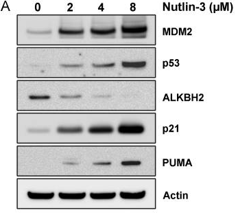
| Western blot | MDM2 / p53 / ALKBH2 / p21 / PUMA RRM1 / RRM2 / p53R2 / p21 / p53 / pS6 / S6 |
|
23258843 | |
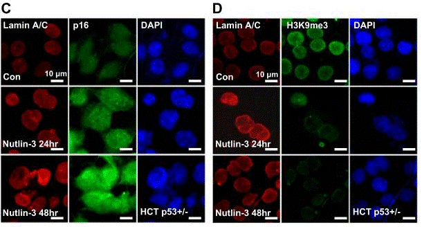
| Immunofluorescence | Lamin A / Lamin C / p16 / H3K9me3 Merlin / cyclin D1 / p53 / MDM2 p53 |
|
30728349 | |
| Growth inhibition assay | Cell viability |
|
29286113 | |
| In Vivo | ||
| In vivo | Oral administration of Nutlin-3 at 200 mg/kg twice daily for 3 weeks significantly inhibits the tumor growth of SJAS-1 xenografts by 90%, comparable with the effect of doxorubicin treatment with 81% inhibition of tumor growth. [1] |
|
|---|---|---|
| Animal Research | Animal Models | Athymic female nude mice (Nu/Nu-nuBR) injected subcutaneously with SJSA-1 cells |
| Dosages | 200 mg/kg | |
| Administration | Orally, twice a day | |
Chemical Information & Solubility
| Molecular Weight | 581.5 | Formula | C30H30Cl2N4O4 |
| CAS No. | 890090-75-2 | SDF | Download Nutlin-3 SDF |
| Smiles | CC(C)OC1=C(C=CC(=C1)OC)C2=NC(C(N2C(=O)N3CCNC(=O)C3)C4=CC=C(C=C4)Cl)C5=CC=C(C=C5)Cl | ||
| Storage (From the date of receipt) | |||
|
In vitro |
DMSO : 100 mg/mL ( (171.96 mM) Moisture-absorbing DMSO reduces solubility. Please use fresh DMSO.) Ethanol : 100 mg/mL Water : Insoluble |
Molecular Weight Calculator |
|
In vivo Add solvents to the product individually and in order. |
In vivo Formulation Calculator |
||||
Preparing Stock Solutions
Molarity Calculator
In vivo Formulation Calculator (Clear solution)
Step 1: Enter information below (Recommended: An additional animal making an allowance for loss during the experiment)
mg/kg
g
μL
Step 2: Enter the in vivo formulation (This is only the calculator, not formulation. Please contact us first if there is no in vivo formulation at the solubility Section.)
% DMSO
%
% Tween 80
% ddH2O
%DMSO
%
Calculation results:
Working concentration: mg/ml;
Method for preparing DMSO master liquid: mg drug pre-dissolved in μL DMSO ( Master liquid concentration mg/mL, Please contact us first if the concentration exceeds the DMSO solubility of the batch of drug. )
Method for preparing in vivo formulation: Take μL DMSO master liquid, next addμL PEG300, mix and clarify, next addμL Tween 80, mix and clarify, next add μL ddH2O, mix and clarify.
Method for preparing in vivo formulation: Take μL DMSO master liquid, next add μL Corn oil, mix and clarify.
Note: 1. Please make sure the liquid is clear before adding the next solvent.
2. Be sure to add the solvent(s) in order. You must ensure that the solution obtained, in the previous addition, is a clear solution before proceeding to add the next solvent. Physical methods such
as vortex, ultrasound or hot water bath can be used to aid dissolving.
Tech Support
Answers to questions you may have can be found in the inhibitor handling instructions. Topics include how to prepare stock solutions, how to store inhibitors, and issues that need special attention for cell-based assays and animal experiments.
Tel: +1-832-582-8158 Ext:3
If you have any other enquiries, please leave a message.
* Indicates a Required Field
Frequently Asked Questions
Question 1:
Is this a racemic mixture of Nutlin-3a and Nutlin-3b or just the Nutlin-3a enantiomer?
Answer:
It is a racemate.
Tags: buy Nutlin-3 | Nutlin-3 supplier | purchase Nutlin-3 | Nutlin-3 cost | Nutlin-3 manufacturer | order Nutlin-3 | Nutlin-3 distributor


