-
 Australia
-
 Austria
-
 Belgium
-
 Brazil
-
 Canada
-
 China
China
-
 Czech Republic
-
 Denmark
-
 Finland
-
 France
-
 Germany
-
 Greece
-
 Hong Kong
-
 Hungary
-
 Iceland
-
 India
India
-
 Ireland
-
 Israel
-
 Italy
Italy
-
 Japan
Japan
-
 Korea
Korea
-
 Luxembourg
-
 Malaysia
-
 Netherlands
-
 New Zealand
-
 Norway
-
 Poland
-
 Qatar
Qatar
-
 Romania
-
 Saudi Arabia
-
 Singapore
-
 Spain
Spain
-
 Sweden
-
 Switzerland
-
 Taiwan
-
 Turkey
-
 United Kingdom
-
 United States
research use only
MK-5108 Aurora Kinase inhibitor
Cat.No.S2770
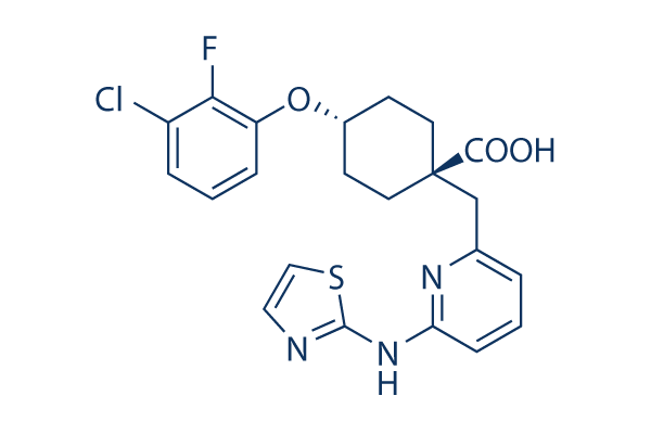
Chemical Structure
Molecular Weight: 461.94
Quality Control
| Related Targets | CDK HSP PD-1/PD-L1 ROCK Wee1 DNA/RNA Synthesis Microtubule Associated Ras KRas Casein Kinase |
|---|---|
| Other Aurora Kinase Inhibitors | Hesperadin Barasertib-HQPA (AZD2811) Alisertib (MLN8237) Tozasertib (VX-680, MK-0457) ZM 447439 MLN8054 Danusertib (PHA-739358) TCS7010 (Aurora A Inhibitor I) AMG-900 CCT137690 |
Cell Culture, Treatment & Working Concentration
| Cell Lines | Assay Type | Concentration | Incubation Time | Formulation | Activity Description | PMID |
|---|---|---|---|---|---|---|
| BL21 (DE3) Rosetta | Function assay | 30 mins | Inhibition of His-tagged human Aurora A kinase (122 to 40 residues) expressed in Escherichia coli BL21 (DE3) Rosetta cells using biotinylated STK2 substrate incubated for 30 mins by HTRF assay, IC50 = 0.000064 μM. | 27391133 | ||
| HeLa Kyoto | Function assay | 20 hrs | Inhibition of Aurora A kinase autophosphorylation at Thr288 in human HeLa Kyoto cells incubated for 20 hrs, IC50 = 0.3 μM. | 27391133 | ||
| HCC1143 | Antiproliferation assay | Antiproliferation activity against human HCC1143 cells assessed as inhibition of cell proliferation, IC50 = 0.42 μM. | 28918096 | |||
| AU565 | Antiproliferation assay | Antiproliferation activity against human AU565 cells assessed as inhibition of cell proliferation, IC50 = 0.45 μM. | 28918096 | |||
| MCF7 | Antiproliferation assay | Antiproliferation activity against human MCF7 cells assessed as inhibition of cell proliferation, IC50 = 0.52 μM. | 28918096 | |||
| HCC1806 | Antiproliferation assay | Antiproliferation activity against human HCC1806 cells assessed as inhibition of cell proliferation, IC50 = 0.56 μM. | 28918096 | |||
| CAL851 | Antiproliferation assay | Antiproliferation activity against human CAL851 cells assessed as inhibition of cell proliferation, IC50 = 0.74 μM. | 28918096 | |||
| HeLa | Function assay | 16 mg/kg | 2 days | Plasma concentration in F344/N Jcl-rnu rat xenografted with luciferase expressing human HeLa cells at 16 mg/kg, po BID for 2 days, Cp = 1.7 μM. | 28918096 | |
| HeLa | Function assay | 32 mg/kg | 2 days | Plasma concentration in F344/N Jcl-rnu rat xenografted with luciferase expressing human HeLa cells at 32 mg/kg, po BID for 2 days, Cp = 4.4 μM. | 28918096 | |
| Saos-2 | qHTS assay | qHTS of pediatric cancer cell lines to identify multiple opportunities for drug repurposing: Primary screen for Saos-2 cells | 29435139 | |||
| MG 63 (6-TG R) | qHTS assay | qHTS of pediatric cancer cell lines to identify multiple opportunities for drug repurposing: Primary screen for MG 63 (6-TG R) cells | 29435139 | |||
| OHS-50 | qHTS assay | qHTS of pediatric cancer cell lines to identify multiple opportunities for drug repurposing: Primary screen for OHS-50 cells | 29435139 | |||
| SK-N-MC | qHTS assay | qHTS of pediatric cancer cell lines to identify multiple opportunities for drug repurposing: Primary screen for SK-N-MC cells | 29435139 | |||
| BL21-Codon-Plus(DE3)-RIL | Function assay | 120 mins | Inhibition of human N-terminal His-tagged Aurora A expressed in Escherichia coli BL21-Codon-Plus(DE3)-RIL cells using 5-carboxy-fluorescein-y-aminobutyric acid-Ala-Leu-Arg-Arg-Ala-Ser-Leu-Gly-NH2 as substrate after 120 mins by fluorescence polarization as, IC50 = 0.00054 μM. | ChEMBL | ||
| HeLaS3 | Cytotoxicity assay | 3 days | Cytotoxicity against human HeLaS3 cells assessed as cell growth inhibition after 3 days by WST-8 assay, IC50 = 1.26 μM. | ChEMBL | ||
| Click to View More Cell Line Experimental Data | ||||||
Solubility
|
In vitro |
DMSO
: 92 mg/mL
(199.16 mM)
Water : Insoluble Ethanol : Insoluble |
Molarity Calculator
|
In vivo |
|||||
In vivo Formulation Calculator (Clear solution)
Step 1: Enter information below (Recommended: An additional animal making an allowance for loss during the experiment)
Step 2: Enter the in vivo formulation (This is only the calculator, not formulation. Please contact us first if there is no in vivo formulation at the solubility Section.)
Calculation results:
Working concentration: mg/ml;
Method for preparing DMSO master liquid: mg drug pre-dissolved in μL DMSO ( Master liquid concentration mg/mL, Please contact us first if the concentration exceeds the DMSO solubility of the batch of drug. )
Method for preparing in vivo formulation: Take μL DMSO master liquid, next addμL PEG300, mix and clarify, next addμL Tween 80, mix and clarify, next add μL ddH2O, mix and clarify.
Method for preparing in vivo formulation: Take μL DMSO master liquid, next add μL Corn oil, mix and clarify.
Note: 1. Please make sure the liquid is clear before adding the next solvent.
2. Be sure to add the solvent(s) in order. You must ensure that the solution obtained, in the previous addition, is a clear solution before proceeding to add the next solvent. Physical methods such
as vortex, ultrasound or hot water bath can be used to aid dissolving.
Chemical Information, Storage & Stability
| Molecular Weight | 461.94 | Formula | C22H21ClFN3O3S |
Storage (From the date of receipt) | |
|---|---|---|---|---|---|
| CAS No. | 1010085-13-8 | Download SDF | Storage of Stock Solutions |
|
|
| Synonyms | VX-689 | Smiles | C1CC(CCC1OC2=C(C(=CC=C2)Cl)F)(CC3=NC(=CC=C3)NC4=NC=CS4)C(=O)O | ||
Mechanism of Action
| Targets/IC50/Ki |
Aurora A
(Cell-free assay) 0.064 nM
|
|---|---|
| In vitro |
MK-5108 inhibits Aurora-A activity in an ATP-competitive manner. This compound shows robust selectivity against the other family kinases Aurora-B (220-fold) and Aurora-C (190-fold) in the biochemical assay. It also reveals high selectivity for Aurora-A over other protein kinases. The compound inhibits only one kinase (TrkA) with <100-fold selectivity. It may be more Aurora-A selective than MLN8054. Consistent with the induction of pHH3-positive cells, this chemical induces accumulation of cells in the G2-M phase. It inhibits the proliferation of tumor cells including HCC1143, AU565, MCF-7, HCC1806 and CAL85-1 with an IC50 of 0.42 μM, 0.45 μM, 0.52 μM, 0.56μM and 0.74 μM, respectively. This compound decreases cell viability in a dose-dependent fashion in all three cell lines including LEIO285, LEIO505 and SK-LSM1 cells with an IC50 of approximately 100 nM. Incubation with it in LEIO285 increases the proportion of cells in G2/M at 48 and 72 hours post-treatment. The compound significant increases in Caspase 3/7 activity when compared to DMSO-treated control cultures at both time points. In LEIO505 cells, it leads to more cells accumulating at G2/M phases at 24 hours but not 48 hours or 72 hours. |
| Kinase Assay |
Biochemical kinase assays
|
|
Recombinant His-tagged human Aurora-A protein is expressed in Escherichia coli and is purified with HisTrap HP column. Purified recombinant human Aurora-B and Aurora-C protein are purchased. Experiments are done in quintuplicate in 96-well plates. The Aurora-A assay reaction is conducted in the presence of 20 μM ATP, 25 μM Tetra-Kemptide [RRR(GLRRASLG)4R-NH2], 1.0 μCi per well [γ-33P]-ATP, 0.1 ng per well Aurora-A in 50 mM Tris-HCl (pH 7.4), 15 mM Mg(OAc)2, and 0.2 mM EDTA at 30°C for 40 minutes. To investigate the inhibition mode of MK-5108 for Aurora-A, the IC50 values of this compound are determined in the presence of different concentrations of ATP. Then, the IC50 value is plotted as a function of ATP concentration to analyze the effect of ATP concentration on the IC50 value of this chemical. The Aurora-B assay reaction is conducted in the presence of 15 μM ATP, 100 μM Kemptide (GLRRASLG-NH2), 1.0 μCi per well [γ-33P]-ATP, 5.0 ng per well Aurora-B in 50 mM Tris-HCl (pH 7.4), 15 mM Mg(OAc)2, and 0.2 mM EDTA at 30 °C for 20 minuts. The Aurora-C assay reaction is conducted in the presence of 40 μM ATP, 100 μM Kemptide, 1.0 μCi per well [γ-33P]-ATP, 15 ng per well Aurora-C in 10 mM MOPS-NaOH (pH 7.4), 5 mM Mg(OAc)2, 1 mM (±) DTT, and 1 mM EGTA at 30°C for 20 minutes. After kinase reactions are terminated by adding 2.0% phosphoric acid, Tetra-Kemptide or Kemptide is trapped on the MultiScreen-PH plate. Wells are washed five times with 0.64% phosphoric acid and then monitored for radioactivity in a liquid scintillation counter.
|
|
| In vivo |
MK-5108 induces pHH3-positive cells at doses of 16 mg/kg and 32 mg/kg. Plasma concentration of this compound at 8 mg/kg and 16 mg/kg are 1.7 μM and 4.4 μM, respectively. This compound treatment results in the induction of pHH3 in tumor and skin tissues, which starts at 2 hours and reachs a maximum at 4 hours. This chemical treatments at 15 mg/kg and 30 mg/kg results in significant tumor growth inhibition with the change in mean tumor volume for the treatment group as a percentage of the mean change in the control group (%T/C) of 10% and −6% at day 11, and 17% and 5% at day 18, respectively. It is well tolerated at both doses, with minimal reduction in body weight. This compound also exhibits significant antitumor activity through intermittent dosing in nude rats bearing SW48 tumors, it at 15 mg/kg and 45 mg/kg causes dose-dependent tumor growth inhibition with a %T/C of 35% and 7% at day 10, and 58% and 32% at day 27, respectively. |
References |
|
Applications
| Methods | Biomarkers | Images | PMID |
|---|---|---|---|
| Western blot | p-Aurora A / Aurora-A / N-MYC |
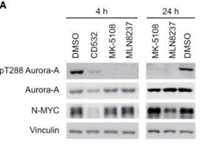
|
29262328 |
| Growth inhibition assay | Cell viability |
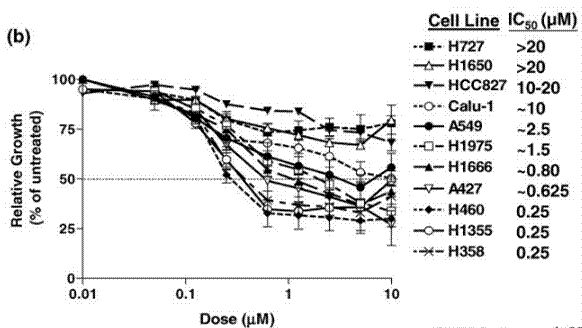
|
24756365 |
Tech Support
Tel: +1-832-582-8158 Ext:3
If you have any other enquiries, please leave a message.
Signaling Pathway Map
Products are for research use only. Not for human use. We do not sell to patients.
©Copyright 2013 Selleck Chemicals. All Rights Reserved.