|
Toll Free: (877) 796-6397 -- USA and Canada only -- |
Fax: +1-832-582-8590 Orders: +1-832-582-8158 |
Tech Support: +1-832-582-8158 Ext:3 Please provide your Order Number in the email. |
Technical Data
| Formula | C24H16F3NO4 |
||||||||||
| Molecular Weight | 439.38 | CAS No. | 393105-53-8 | ||||||||
| Solubility (25°C)* | In vitro | DMSO | 88 mg/mL (200.28 mM) | ||||||||
| Ethanol | 22 mg/mL (50.07 mM) | ||||||||||
| Water | Insoluble | ||||||||||
| In vivo (Add solvents to the product individually and in order) |
|
||||||||||
|
* <1 mg/ml means slightly soluble or insoluble. * Please note that Selleck tests the solubility of all compounds in-house, and the actual solubility may differ slightly from published values. This is normal and is due to slight batch-to-batch variations. * Room temperature shipping (Stability testing shows this product can be shipped without any cooling measures.) |
|||||||||||
Preparing Stock Solutions
Biological Activity
| Description | Tiplaxtinin(PAI-039) is an orally efficacious and selective plasminogen activator inhibitor-1 (PAI-1) inhibitor with IC50 of 2.7 μM. | ||
|---|---|---|---|
| Targets |
|
||
| In vitro | In a panel of human bladder cell lines, PAI-1 results in the reduction of cellular proliferation, cell adhesion, and colony formation, and the induction of apoptosis and anoikis. [4] | ||
| In vivo | In a rat carotid thrombosis model, Tiplaxtinin (1 mg/kg, p.o.) increases time to occlusion and prevents the carotid blood flow reduction. [1] In C57BL/6J mice, (1 mg/g chow) attenuates Ang II-induced aortic remodeling. [2] In untreated type 1 diabetic mice, Tiplaxtinin (p.o.) restores skeletal muscle regeneration. [3] In athymic mice bearing human cancer cell line T24 and HeLa xenografts, Tiplaxtinin (1 mg/kg, p.o.) reduces tumor xenograft growth, associated with a reduction in tumor angiogenesis, a reduction in cellular proliferation, and an increase in apoptosis. [4] |
Protocol (from reference)
| Kinase Assay:[1] |
|
|---|---|
| Cell Assay:[4] |
|
| Animal Study:[1] |
|
References
Customer Product Validation

-
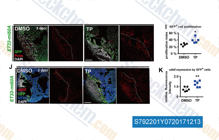
Data from [Data independently produced by , , Development, 2017, 144(8):1425-1440]
Selleck's Tiplaxtinin (PAI-039) has been cited by 16 publications
| ProBDNF as a Myokine in Skeletal Muscle Injury: Role in Inflammation and Potential for Therapeutic Modulation of p75NTR [ Int J Mol Sci, 2025, 26(1)401] | PubMed: 39796256 |
| hapln1a+ cells guide coronary growth during heart morphogenesis and regeneration [ Nat Commun, 2023, 14(1):3505] | PubMed: 37311876 |
| hapln1a+ cells guide coronary growth during heart morphogenesis and regeneration [ Nat Commun, 2023, 14(1):3505] | PubMed: 37311876 |
| CSE reduces OTUD4 triggering lung epithelial cell apoptosis via PAI-1 degradation [ Cell Death Dis, 2023, 14(9):614] | PubMed: 37726265 |
| CSE reduces OTUD4 triggering lung epithelial cell apoptosis via PAI-1 degradation [ Cell Death Dis, 2023, 10.1038/s41419-023-06131-1] | PubMed: 37726265 |
| Ligand-mediated PAI-1 inhibition in a mouse model of peritoneal carcinomatosis [ Cell Rep Med, 2022, 3(2):100526] | PubMed: 35243423 |
| ProBDNF Dependence of LTD and Fear Extinction Learning in the Amygdala of Adult Mice [ Cereb Cortex, 2022, 32(7):1350-1364] | PubMed: 34470044 |
| Development of potent and selective inhibitors targeting the papain-like protease of SARS-CoV-2 [ Cell Chem Biol, 2021, S2451-9456(21)00213-0] | PubMed: 33979649 |
| Cancer‑associated fibroblast‑induced M2‑polarized macrophages promote hepatocellular carcinoma progression via the plasminogen activator inhibitor‑1 pathway [ Int J Oncol, 2021, 59(2)59] | PubMed: 34195849 |
| Long-term depression at hippocampal mossy fiber-CA3 synapses involves BDNF but is not mediated by p75NTR signaling [ Sci Rep, 2021, 11(1):8535] | PubMed: 33879805 |
RETURN POLICY
Selleck Chemical’s Unconditional Return Policy ensures a smooth online shopping experience for our customers. If you are in any way unsatisfied with your purchase, you may return any item(s) within 7 days of receiving it. In the event of product quality issues, either protocol related or product related problems, you may return any item(s) within 365 days from the original purchase date. Please follow the instructions below when returning products.
SHIPPING AND STORAGE
Selleck products are transported at room temperature. If you receive the product at room temperature, please rest assured, the Selleck Quality Inspection Department has conducted experiments to verify that the normal temperature placement of one month will not affect the biological activity of powder products. After collecting, please store the product according to the requirements described in the datasheet. Most Selleck products are stable under the recommended conditions.
NOT FOR HUMAN, VETERINARY DIAGNOSTIC OR THERAPEUTIC USE.
