|
Toll Free: (877) 796-6397 -- USA and Canada only -- |
Fax: +1-832-582-8590 Orders: +1-832-582-8158 |
Tech Support: +1-832-582-8158 Ext:3 Please provide your Order Number in the email. |
Technical Data
| Formula | C25H17F2N5O3S |
|||
| Molecular Weight | 505.5 | CAS No. | 1086062-66-9 | |
| Solubility (25°C)* | In vitro | DMSO | 101 mg/mL (199.8 mM) | |
| Water | Insoluble | |||
| Ethanol | Insoluble | |||
|
* <1 mg/ml means slightly soluble or insoluble. * Please note that Selleck tests the solubility of all compounds in-house, and the actual solubility may differ slightly from published values. This is normal and is due to slight batch-to-batch variations. * Room temperature shipping (Stability testing shows this product can be shipped without any cooling measures.) |
||||
Preparing Stock Solutions
Biological Activity
| Description | Omipalisib (GSK2126458, GSK458) is a highly selective and potent inhibitor of p110α/β/δ/γ, mTORC1/2 with Ki of 0.019 nM/0.13 nM/0.024 nM/0.06 nM and 0.18 nM/0.3 nM in cell-free assays, respectively. Omipalisib induces autophagy. Phase 1. | |||||||||||
|---|---|---|---|---|---|---|---|---|---|---|---|---|
| Targets |
|
|||||||||||
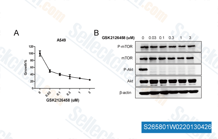
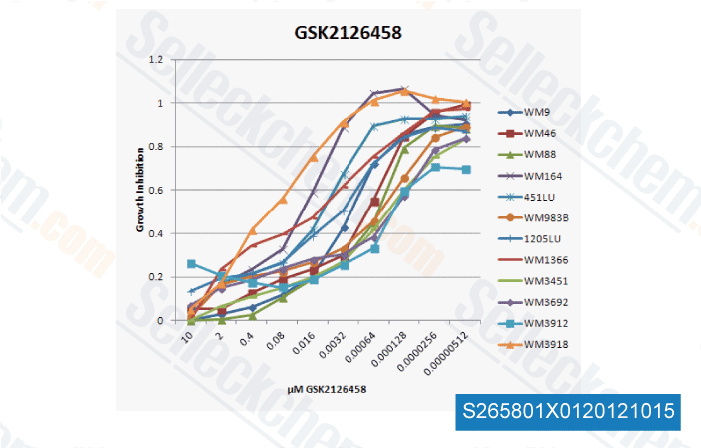
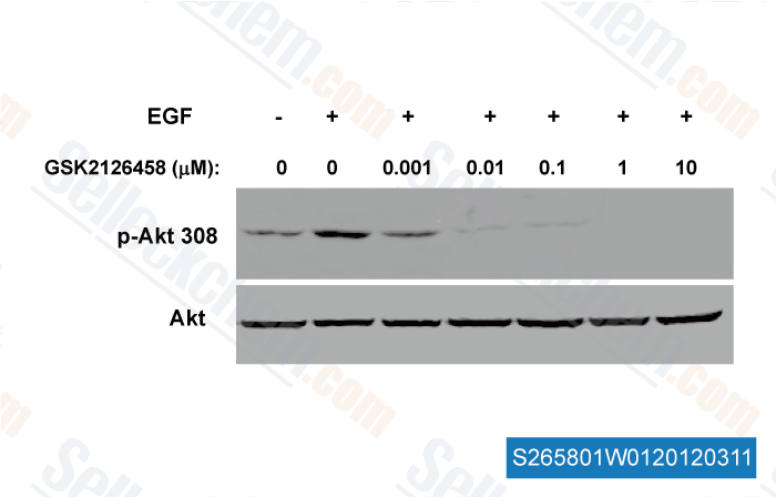
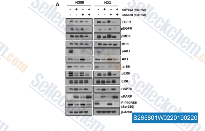
| In vitro | GSK2126458 potently inhibits the activity of common activating mutants of p110α (E542K, E545K, and H1047R) found in human cancer with Ki of 8 pM, 8 pM and 9 pM, respectively. [1] GSK2126458 causes a significant reduction in the levels of pAkt-S473 with remarkable potency in T47D and BT474 cells with IC50 of 0.41 nM and 0.18 nM, respectively. Furthermore, GSK2126458 leads to a G1 cell cycle arrest and produces the inhibitory effect on cell proliferation in a large panel of cell lines, including T47D and BT474 breast cancer lines with IC50 of 3 nM and 2.4 nM, respectively. [1] | |||||||||||
| In vivo | In a BT474 human tumor xenograft model, GSK2126458 treatment results in a dose-dependent reduction in pAkt-S473 levels, and exhibited dose-dependent tumor growth inhibition at a low dose of 300 μg /kg. Besides, GSK2126458 shows low blood clearance and good oral bioavailability in four preclinical species (mouse, rat, dog, and monkey). [1] |
Protocol (from reference)
| Kinase Assay:[1] |
|
|---|---|
| Cell Assay:[1] |
|
| Animal Study:[1] |
|
References
|
Customer Product Validation

-
Data independently produced by Dr.Wang Wei from NanFang Hospital,

-
, , One customer

-
, , Dr. Zhang of Tianjin Medical University

-
Data from [Data independently produced by , , Cancer Lett, 2017, 406:47-53]
Selleck's Omipalisib (GSK2126458) has been cited by 63 publications
| Trametinib sensitizes KRAS-mutant lung adenocarcinoma tumors to PD-1/PD-L1 axis blockade via Id1 downregulation [ Mol Cancer, 2024, 23(1):78] | PubMed: 38643157 |
| Trametinib sensitizes KRAS-mutant lung adenocarcinoma tumors to PD-1/PD-L1 axis blockade via Id1 downregulation [ Mol Cancer, 2024, 23(1):78] | PubMed: 38643157 |
| NKX2-5 regulates vessel remodeling in scleroderma-associated pulmonary arterial hypertension [ JCI Insight, 2024, 9(10)e164191] | PubMed: 38652537 |
| Characterizing heterogeneous single-cell dose responses computationally and experimentally using threshold inhibition surfaces and dose-titration assays [ NPJ Syst Biol Appl, 2024, 10(1):42] | PubMed: 38637530 |
| Analysis of hsa_circ_0136256 as a biomarker for fibrosis in systemic sclerosis [ BMC Biotechnol, 2024, 24(1):91] | PubMed: 39538329 |
| "Proteotranscriptomic analysis of advanced colorectal cancer patient derived organoids for drug sensitivity prediction" [ J Exp Clin Cancer Res, 2023, 42(1):8] | PubMed: 36604765 |
| PI3K/mTOR inhibitors promote G6PD autophagic degradation and exacerbate oxidative stress damage to radiosensitize small cell lung cancer [ Cell Death Dis, 2023, 14(10):652] | PubMed: 37802999 |
| PI3K/mTOR inhibitors promote G6PD autophagic degradation and exacerbate oxidative stress damage to radiosensitize small cell lung cancer [ Cell Death Dis, 2023, 14(10):652] | PubMed: 37802999 |
| Integrin-mediated electric axon guidance underlying optic nerve formation in the embryonic chick retina [ Commun Biol, 2023, 6(1):680] | PubMed: 37391492 |
| Integrin-mediated electric axon guidance underlying optic nerve formation in the embryonic chick retina [ Commun Biol, 2023, 6(1):680] | PubMed: 37391492 |
RETURN POLICY
Selleck Chemical’s Unconditional Return Policy ensures a smooth online shopping experience for our customers. If you are in any way unsatisfied with your purchase, you may return any item(s) within 7 days of receiving it. In the event of product quality issues, either protocol related or product related problems, you may return any item(s) within 365 days from the original purchase date. Please follow the instructions below when returning products.
SHIPPING AND STORAGE
Selleck products are transported at room temperature. If you receive the product at room temperature, please rest assured, the Selleck Quality Inspection Department has conducted experiments to verify that the normal temperature placement of one month will not affect the biological activity of powder products. After collecting, please store the product according to the requirements described in the datasheet. Most Selleck products are stable under the recommended conditions.
NOT FOR HUMAN, VETERINARY DIAGNOSTIC OR THERAPEUTIC USE.
