|
How to Cite 1. For In-Text Citation (Materials & Methods): 2. For Key Resources Table: |
||
|
Toll Free: (877) 796-6397 -- USA and Canada only -- |
Fax: +1-832-582-8590 Orders: +1-832-582-8158 |
Tech Support: +1-832-582-8158 Ext:3 Please provide your Order Number in the email. We strive to reply to |
Technical Data
| Formula | C19H21F2N7O2 |
||||||
| Molecular Weight | 417.41 | CAS No. | 475110-96-4 | ||||
| Solubility (25°C)* | In vitro | DMSO | 21 mg/mL (50.31 mM) | ||||
| Water | Insoluble | ||||||
| Ethanol | Insoluble | ||||||
| In vivo (Add solvents to the product individually and in order) |
|
||||||
|
* <1 mg/ml means slightly soluble or insoluble. * Please note that Selleck tests the solubility of all compounds in-house, and the actual solubility may differ slightly from published values. This is normal and is due to slight batch-to-batch variations. * Room temperature shipping (Stability testing shows this product can be shipped without any cooling measures.) |
|||||||
Preparing Stock Solutions
Biological Activity
| Description | ZSTK474 inhibits class I PI3K isoforms with IC50 of 37 nM in a cell-free assay, mostly PI3Kδ. Phase1/2. | ||||||||||
|---|---|---|---|---|---|---|---|---|---|---|---|
| Targets |
|
||||||||||
| In vitro | ZSTK474 at 1 μM potently reduces PI3K activity to 4.7% of the control level, whereas LY2194002 only reduces the activity to 44.6% of the control. This compound inhibits the activities of recombinant p110β, -γ, and -δ with IC50 of 17 nM, 53 nM, and 6 nM, respectively. It shows potent antiproliferative activity against a panel of 39 human cancer cell lines with mean GI50 of 0.32 μM, more effectively than that of LY294002 or wortmannin with mean GI50 of 7.4 μM or 10 μM, respectively. This chemical treatment at 1 μM blocks membrane ruffling and generation of PIP3 induced by platelet-derived growth factor in murine embryonic fibroblasts (MEFs). It at 10 μM induces apoptosis in OVCAR3 cells, and induces complete G1-phase arrest but not apoptosis in A549 cells. This compound treatment at 0.5 μM significantly decreases the level of phosphorylated Akt and GSK-3β, as well as the cyclin D1 protein expression. It also inhibits the phosphorylation of other downstream signaling components that are involved in regulating cell proliferation including FKHRL1, FKHR, TSC-2, mTOR, and p70S6K in a dose-dependent manner. [1] This chemical does not inhibit mTOR at 0.1 μM, and even at a concentration of 100 μM, it inhibits mTOR activity less than 40%. [2] It blocks VEGF-induced cell migration and the tube formation in human umbilical vein endothelial cells (HUVECs), and inhibits the expression of HIF-1α and secretion of VEGF in RXF-631L cells, exhibiting potent in vitro antiangiogenic activity. [3] This compound treatment inhibits the production of IFNγ and IL-17 in concanavalin A-activated T cells, and inhibits the proliferation and PGE(2) production by fibroblast-like synovial cells (FLS). [6] |
||||||||||
| In vivo | Oral administration of ZSTK474 inhibits the growth of subcutaneously implanted mouse B16F10 melanoma tumors in a dose-dependent manner, producing tumor regression of 28.5%, 7.1%, or 4.9% on day 14 at 100, 200, or 400 mg/kg, respectively, which is superior to that of the four major anticancer drugs at their respective maximum tolerable doses with tumor regression of 96%, 35.7%, 24%, or 68.3%, respectively. This compound treatment at 400 mg/kg completely inhibits the growth of A549, PC-3, and WiDr xenografts in mice, and induces the regression of A549 xenograft tumors. [1] It significantly inhibits tumor growth in the RXF-631L xenograft model, correlated with a significantly reduced number of microvessels in the treated mice. [3] Oral administration of this chemical ameliorates the progression of adjuvant-induced arthritis (AIA) in rats. [6] | ||||||||||
| Features | First orally administered PI3K inhibitor used in vivo. |
Protocol (from reference)
| Kinase Assay: |
|
|---|---|
| Cell Assay: |
|
| Animal Study: |
|
References
|
Customer Product Validation

-
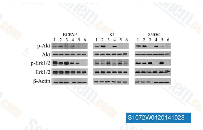
Data from [ Invest New Drugs , 2014 , 32(4), 626-35 ]

-
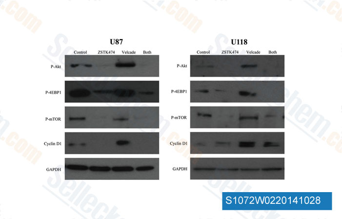
Data from [ Int J Oncol , 2014 , 44(2), 557-62 ]

-
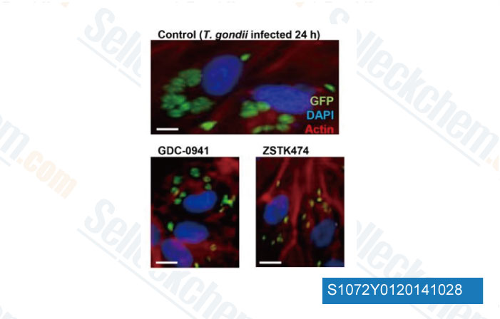
Data from [ PLoS One , 2013 , 8(6), e66306 ]

-
, , Saraswati Sukumar of Johns Hopkins University School of Medicine
Selleck's ZSTK474 Has Been Cited by 88 Publications
| PI3K-Akt signalling regulates Scx-lineage tenocytes and Tppp3-lineage paratenon sheath cells in neonatal tendon regeneration [ Nat Commun, 2025, 16(1):3734] | PubMed: 40254618 |
| Perinatal thymic-derived CD8αβ-expressing γδ T cells are innate IFN-γ producers that expand in IL-7R-STAT5B-driven neoplasms [ Nat Immunol, 2024, 10.1038/s41590-024-01855-4] | PubMed: 38802512 |
| Perinatal thymic-derived CD8αβ-expressing γδ T cells are innate IFN-γ producers that expand in IL-7R-STAT5B-driven neoplasms [ Nat Immunol, 2024, 25(7):1207-1217] | PubMed: 38802512 |
| Reporter cell lines to screen for inhibitors or regulators of the KRAS-RAF-MEK1/2-ERK1/2 pathway [ Biochem J, 2024, 481(6):405-422] | PubMed: 38381045 |
| B cell adapter for PI 3-kinase (BCAP) coordinates antigen internalization and trafficking through the B cell receptor [ Sci Adv, 2024, 10(46):eadp1747] | PubMed: 39546610 |
| Multiplex single-cell chemical genomics reveals the kinase dependence of the response to targeted therapy [ Cell Genom, 2024, 4(2):100487] | PubMed: 38278156 |
| Stellettin B Sensitizes Glioblastoma to DNA-Damaging Treatments by Suppressing PI3K-Mediated Homologous Recombination Repair [ Adv Sci (Weinh), 2023, 10(3):e2205529] | PubMed: 36453577 |
| Young and undamaged recombinant albumin alleviates T2DM by improving hepatic glycolysis through EGFR and protecting islet β cells in mice [ J Transl Med, 2023, 21(1):89] | PubMed: 36747238 |
| Young and undamaged recombinant albumin alleviates T2DM by improving hepatic glycolysis through EGFR and protecting islet β cells in mice [ J Transl Med, 2023, 21(1):89] | PubMed: 36747238 |
| HDAC Inhibition Restores Response to HER2-Targeted Therapy in Breast Cancer via PHLDA1 Induction [ Int J Mol Sci, 2023, 24(7)6228] | PubMed: 37047202 |
RETURN POLICY
Selleck Chemical’s Unconditional Return Policy ensures a smooth online shopping experience for our customers. If you are in any way unsatisfied with your purchase, you may return any item(s) within 7 days of receiving it. In the event of product quality issues, either protocol related or product related problems, you may return any item(s) within 365 days from the original purchase date. Please follow the instructions below when returning products.
SHIPPING AND STORAGE
Selleck products are transported at room temperature. If you receive the product at room temperature, please rest assured, the Selleck Quality Inspection Department has conducted experiments to verify that the normal temperature placement of one month will not affect the biological activity of powder products. After collecting, please store the product according to the requirements described in the datasheet. Most Selleck products are stable under the recommended conditions.
NOT FOR HUMAN, VETERINARY DIAGNOSTIC OR THERAPEUTIC USE.
