|
Toll Free: (877) 796-6397 -- USA and Canada only -- |
Fax: +1-832-582-8590 Orders: +1-832-582-8158 |
Tech Support: +1-832-582-8158 Ext:3 Please provide your Order Number in the email. |
Technical Data
| Formula | C10H12FN5O4 |
||||||
| Molecular Weight | 285.23 | CAS No. | 21679-14-1 | ||||
| Solubility (25°C)* | In vitro | DMSO | 57 mg/mL (199.83 mM) | ||||
| Water | Insoluble | ||||||
| Ethanol | Insoluble | ||||||
| In vivo (Add solvents to the product individually and in order) |
|
||||||
|
* <1 mg/ml means slightly soluble or insoluble. * Please note that Selleck tests the solubility of all compounds in-house, and the actual solubility may differ slightly from published values. This is normal and is due to slight batch-to-batch variations. * Room temperature shipping (Stability testing shows this product can be shipped without any cooling measures.) |
|||||||
Preparing Stock Solutions
Biological Activity
| Description | Fludarabine is a STAT1 activation inhibitor which causes a specific depletion of STAT1 protein (and mRNA) but not of other STATs. Also a DNA synthesis inhibitor in vascular smooth muscle cells. Fludarabine induces apoptosis. | |
|---|---|---|
| Targets |
|
|
| In vitro | Fludarabine efficiently inhibits the proliferation of RPMI 8226 cells with IC50 of 1.54 μg/mL. The IC50 of Fludarabine against MM.1S and MM.1R cells is 13.48 μg/mL and 33.79 μg/mL, respectively. In contrast, U266 cells are resistant to Fludarabine with IC50 of 222.2 μg/mL. Fludarabine treatment results in increased number of cells in the G1 phase of cell cycle, accompanied with a concomitant reduction of cells at the S phase of cell cycle in a time-dependent manner. Fludarabine induces a cell cycle block and triggers apoptosis in MM cells. Fludarabine triggers time-dependent cleavage of caspase-8, -9, and -3, -7, followed by PARP cleavage. Fludarabine increases expression of Bax in a time-dependent fashion, while the expression of Bak doesn't change. After exposure to Fludarabine for 12 hours, RPMI 8226 cells shows a loss of membrane potential with 61.05% of the cells expressing low fluorescence of rhodamine 123 compared with 8.62% of cells in untreated control. [1] To enhance solubility, Fludarabine is formulated as the monophosphate (F-ara-AMP, fudarabine), which is instantaneously and quantitatively dephosphorylated to the parent nucleoside upon intravenous infusion. Inside the cells rephosphorylation occurs which leads to fuoroadenine arabinoside triphosphate (F-ara-ATP), the major cytotoxic metabolite of F-ara-A. [2] Fludarabine can also induce pro-inflammatory stimulation of monocytic cells, as evaluated by increased expression of ICAM-1 and IL-8 release. [3] Fludarabine does not affect the growth of ovarian cancer cell lines, whereas it induces marked and dose-dependent inhibition of proliferation in melanoma cell lines. [4] Fludarabine is an inhibitor of STAT1 that specifically reduces STAT1 without affecting other STAT family members[5]. In addition to cytoplasmic accumulation, repeated low-dose cisplatin (RLDC) induces HMGB1 expression, which is marked suppressed by STAT1 knockdown. Consistently, fludarabine suppresses HMGB1 expression during RLDC treatment dose-dependently in RLDC-treat renal tubular cells[5]. |
|
| In vivo | Tumors treated with PBS grow rapidly to approx-imately 10-fold their initial volume in 25 day, whereas, the tumors in the Fludarabine at 40 mg/kg increase less than 5-fold. A significant antitumor effect of 40 mg/kg Fludarabine on RPMI8226 tumor growth is demonstrated. RPMI8226 tumors treated with 40 mg/kg Fludarabine at day 10 increase apoptotic nuclei. Fludarabine is effective in suppressing RPMI8226 myeloma xenografts in SCID mice. [1] |
Protocol (from reference)
| Cell Assay: |
|
|---|---|
| Animal Study: |
|
References
Customer Product Validation

-
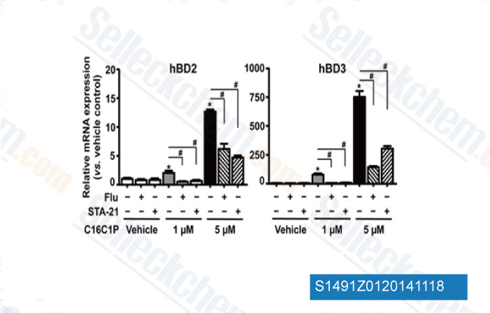
Data from [Data independently produced by Mol Cell Biol, 2014, 34(24), 4368-78.]

-
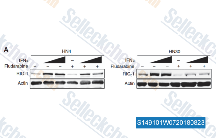
Data from [Data independently produced by , , Br J Cancer, 2018, 118(4):509-521]

-
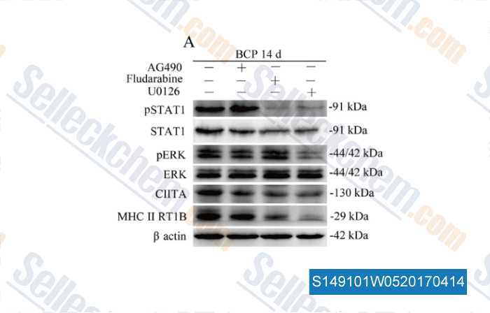
Data from [Data independently produced by , , Brain Behav Immun, 2017, 60:161-173]

-
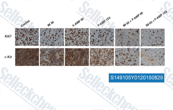
Data from [Data independently produced by , , Mol Cancer Ther, 2014, 13(10): 2276-87 ]
Selleck's Fludarabine has been cited by 211 publications
| MSC transplantation ameliorates depression in lupus by suppressing Th1 cell-shaped synaptic stripping [ JCI Insight, 2025, e181885] | PubMed: 40048256 |
| Adenosine deaminase and deoxyadenosine regulate intracellular immune response in C. elegans [ iScience, 2025, 28(3):111950] | PubMed: 40034845 |
| NAT10-mediated mRNA N4-acetylcytidine reprograms serine metabolism to drive leukaemogenesis and stemness in acute myeloid leukaemia [ Nat Cell Biol, 2024, ] | PubMed: 39506072 |
| Type 1 interferons and Foxo1 down-regulation play a key role in age-related T-cell exhaustion in mice [ Nat Commun, 2024, 15(1):1718] | PubMed: 38409097 |
| Succinate dehydrogenase deficiency-driven succinate accumulation induces drug resistance in acute myeloid leukemia via ubiquitin-cullin regulation [ Nat Commun, 2024, 15(1):9820] | PubMed: 39537588 |
| PD-L1-expressing tumor-associated macrophages are immunostimulatory and associate with good clinical outcome in human breast cancer [ Cell Rep Med, 2024, 5(2):101420] | PubMed: 38382468 |
| The involvement of the Stat1/Nrf2 pathway in exacerbating Crizotinib-induced liver injury: implications for ferroptosis [ Cell Death Dis, 2024, 15(8):600] | PubMed: 39160159 |
| Lnc-CCNH-8 promotes immune escape by up-regulating PD-L1 in hepatocellular carcinoma [ Mol Ther Nucleic Acids, 2024, 35(1):102125] | PubMed: 38356866 |
| The E3 ubiquitin ligase Herc1 modulates the response to nucleoside analogs in acute myeloid leukemia [ Blood Adv, 2024, 8(20):5315-5329] | PubMed: 39093953 |
| Intracellular osteopontin potentiates the immunosuppressive activity of mesenchymal stromal cells [ Stem Cell Res Ther, 2024, 15(1):366] | PubMed: 39407354 |
RETURN POLICY
Selleck Chemical’s Unconditional Return Policy ensures a smooth online shopping experience for our customers. If you are in any way unsatisfied with your purchase, you may return any item(s) within 7 days of receiving it. In the event of product quality issues, either protocol related or product related problems, you may return any item(s) within 365 days from the original purchase date. Please follow the instructions below when returning products.
SHIPPING AND STORAGE
Selleck products are transported at room temperature. If you receive the product at room temperature, please rest assured, the Selleck Quality Inspection Department has conducted experiments to verify that the normal temperature placement of one month will not affect the biological activity of powder products. After collecting, please store the product according to the requirements described in the datasheet. Most Selleck products are stable under the recommended conditions.
NOT FOR HUMAN, VETERINARY DIAGNOSTIC OR THERAPEUTIC USE.
