|
Toll Free: (877) 796-6397 -- USA and Canada only -- |
Fax: +1-832-582-8590 Orders: +1-832-582-8158 |
Tech Support: +1-832-582-8158 Ext:3 Please provide your Order Number in the email. |
Technical Data
| Formula | C35H58O9 |
|||
| Molecular Weight | 622.83 | CAS No. | 88899-55-2 | |
| Solubility (25°C)* | In vitro | DMSO | 100 mg/mL (160.55 mM) | |
| Water | Insoluble | |||
| Ethanol | Insoluble | |||
|
* <1 mg/ml means slightly soluble or insoluble. * Please note that Selleck tests the solubility of all compounds in-house, and the actual solubility may differ slightly from published values. This is normal and is due to slight batch-to-batch variations. * Room temperature shipping (Stability testing shows this product can be shipped without any cooling measures.) |
||||
Preparing Stock Solutions
Biological Activity
| Description | Bafilomycin A1(Baf-A1) is a vacuolar H+-ATPase inhibitor with IC50 of 0.44 nM. Bafilomycin A1 is found to inhibit autophagy while induces apoptosis. | ||
|---|---|---|---|
| Targets |
|
||
| In vitro | Bafilomycin A1 is a toxic macrolide antibiotic derived from Streptomyces griseus. Bafilomycin A1 inhibits the short circuit current induced by the outer mantle epithelium (OME). The IC50 and maximum inhibition dose of Bafilomycin A1 are 0.17 μM and 0.5 μM, respectively. [2] In addition, Bilomycin A1 inhibits the acid influx with an IC50 value of 0.4 nM. Bafilomycin A1 inhibits the acidification dose-dependently resulting in a lower quenching, and thus a higher fluorescence. [3] Bafilomycin A1 prevents the vacuolization of Hela cells induced by H. pylori, with an inhibitory concentration giving 50% of maximal (ID50) of 4 nM. Bafilomycin A1 is also very efficient in restoring vacuolated cells to a normal appearance. [4] Bafilomycin A1 also affects the transport of endocytosed material from early to late endocytic compartments. Bafilomycin not only dissipates the low endosomal pH but also blocks transport from early to late endosomes in HeLa cells. [5] Bafilomycin A1 at doses of 0.1-1 μM completely inhibits the acidification of lysosomes revealed by the incubation with acridine orange in BNL CL.2 and A431 cells. [6] When Bafilomycin A1 is added to Hanks' balanced salt solution, endogenous protein degradation is strongly inhibited and numerous autophagosomes accumulated in H-4-II-E cells. Bafilomycin A1 also prevents the appearance of endocytosed HRP in autophagic vacuoles. [7] | ||
| In vivo | Bafilomycin A1 (1 μM and 0.1 μM) completely inhibits the resorptive activity of cultured osteoclasts. [8] Bafilomycin A1 dose-dependently inhibits the rate of Na+ uptake in young tilapia with a Ki of 0.16 μM. [9] |
Protocol (from reference)
| Kinase Assay: |
|
|---|---|
| Cell Assay: |
|
| Animal Study: |
|
References
Customer Product Validation

-
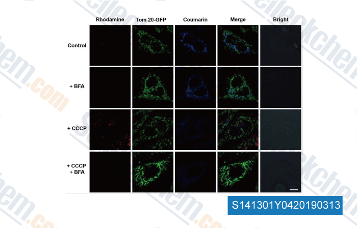
Data from [Data independently produced by , , Chem Sci, 2017, 8(3):1915-1921]

-
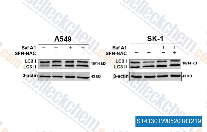
Data from [Data independently produced by , , Cancer Lett, 2018, 431:85-95]

-
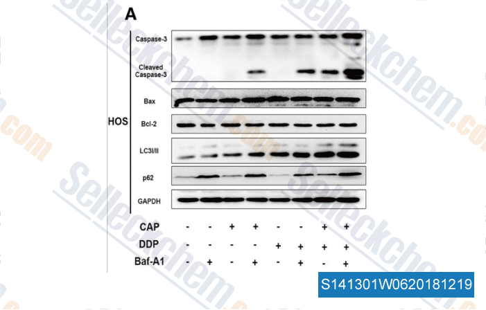
Data from [Data independently produced by , , J Exp Clin Cancer Res, 2018, 37(1):251]

-
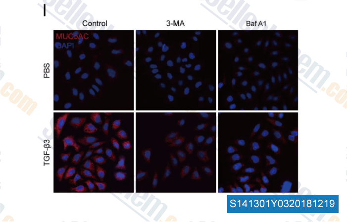
Data from [Data independently produced by , , EBioMedicine, 2018, 33:242-252]
Selleck's Bafilomycin A1 (Baf-A1) has been cited by 545 publications
| Oncogenic RAS induces a distinctive form of non-canonical autophagy mediated by the P38-ULK1-PI4KB axis [ Cell Res, 2025, 10.1038/s41422-025-01085-9] | PubMed: 40055523 |
| Host FSTL1 defines the impact of stem cell therapy on liver fibrosis by potentiating the early recruitment of inflammatory macrophages [ Signal Transduct Target Ther, 2025, 10(1):81] | PubMed: 40050288 |
| Alterations in PD-L1 succinylation shape anti-tumor immune responses in melanoma [ Nat Genet, 2025, 57(3):680-693] | PubMed: 40069506 |
| ACSS2 drives senescence-associated secretory phenotype by limiting purine biosynthesis through PAICS acetylation [ Nat Commun, 2025, 16(1):2071] | PubMed: 40021646 |
| The O-glycosyltransferase C1GALT1 promotes EWSR1::FLI1 expression and is a therapeutic target for Ewing sarcoma [ Nat Commun, 2025, 16(1):1267] | PubMed: 39894896 |
| S100P is a ferroptosis suppressor to facilitate hepatocellular carcinoma development by rewiring lipid metabolism [ Nat Commun, 2025, 16(1):509] | PubMed: 39779666 |
| Extracellular Histones as Exosome Membrane Proteins Regulated by Cell Stress [ J Extracell Vesicles, 2025, 14(2):e70042] | PubMed: 39976275 |
| NEAT1-mediated regulation of proteostasis and mRNA localization impacts autophagy dysregulation in Rett syndrome [ Nucleic Acids Res, 2025, 53(4)gkaf074] | PubMed: 39921568 |
| Disrupting AGR2/IGF1 paracrine and reciprocal signaling for pancreatic cancer therapy [ Cell Rep Med, 2025, 6(2):101927] | PubMed: 39914384 |
| A genome-wide association study identified PRKCB as a causal gene and therapeutic target for Mycobacterium avium complex disease [ Cell Rep Med, 2025, S2666-3791(24)00694-3] | PubMed: 39848245 |
RETURN POLICY
Selleck Chemical’s Unconditional Return Policy ensures a smooth online shopping experience for our customers. If you are in any way unsatisfied with your purchase, you may return any item(s) within 7 days of receiving it. In the event of product quality issues, either protocol related or product related problems, you may return any item(s) within 365 days from the original purchase date. Please follow the instructions below when returning products.
SHIPPING AND STORAGE
Selleck products are transported at room temperature. If you receive the product at room temperature, please rest assured, the Selleck Quality Inspection Department has conducted experiments to verify that the normal temperature placement of one month will not affect the biological activity of powder products. After collecting, please store the product according to the requirements described in the datasheet. Most Selleck products are stable under the recommended conditions.
NOT FOR HUMAN, VETERINARY DIAGNOSTIC OR THERAPEUTIC USE.
