- Bioactive Compounds
- By Signaling Pathways
- PI3K/Akt/mTOR
- Epigenetics
- Methylation
- Immunology & Inflammation
- Protein Tyrosine Kinase
- Angiogenesis
- Apoptosis
- Autophagy
- ER stress & UPR
- JAK/STAT
- MAPK
- Cytoskeletal Signaling
- Cell Cycle
- TGF-beta/Smad
- DNA Damage/DNA Repair
- Compound Libraries
- Popular Compound Libraries
- Customize Library
- Clinical and FDA-approved Related
- Bioactive Compound Libraries
- Inhibitor Related
- Natural Product Related
- Metabolism Related
- Cell Death Related
- By Signaling Pathway
- By Disease
- Anti-infection and Antiviral Related
- Neuronal and Immunology Related
- Fragment and Covalent Related
- FDA-approved Drug Library
- FDA-approved & Passed Phase I Drug Library
- Preclinical/Clinical Compound Library
- Bioactive Compound Library-I
- Bioactive Compound Library-Ⅱ
- Kinase Inhibitor Library
- Express-Pick Library
- Natural Product Library
- Human Endogenous Metabolite Compound Library
- Alkaloid Compound LibraryNew
- Angiogenesis Related compound Library
- Anti-Aging Compound Library
- Anti-alzheimer Disease Compound Library
- Antibiotics compound Library
- Anti-cancer Compound Library
- Anti-cancer Compound Library-Ⅱ
- Anti-cancer Metabolism Compound Library
- Anti-Cardiovascular Disease Compound Library
- Anti-diabetic Compound Library
- Anti-infection Compound Library
- Antioxidant Compound Library
- Anti-parasitic Compound Library
- Antiviral Compound Library
- Apoptosis Compound Library
- Autophagy Compound Library
- Calcium Channel Blocker LibraryNew
- Cambridge Cancer Compound Library
- Carbohydrate Metabolism Compound LibraryNew
- Cell Cycle compound library
- CNS-Penetrant Compound Library
- Covalent Inhibitor Library
- Cytokine Inhibitor LibraryNew
- Cytoskeletal Signaling Pathway Compound Library
- DNA Damage/DNA Repair compound Library
- Drug-like Compound Library
- Endoplasmic Reticulum Stress Compound Library
- Epigenetics Compound Library
- Exosome Secretion Related Compound LibraryNew
- FDA-approved Anticancer Drug LibraryNew
- Ferroptosis Compound Library
- Flavonoid Compound Library
- Fragment Library
- Glutamine Metabolism Compound Library
- Glycolysis Compound Library
- GPCR Compound Library
- Gut Microbial Metabolite Library
- HIF-1 Signaling Pathway Compound Library
- Highly Selective Inhibitor Library
- Histone modification compound library
- HTS Library for Drug Discovery
- Human Hormone Related Compound LibraryNew
- Human Transcription Factor Compound LibraryNew
- Immunology/Inflammation Compound Library
- Inhibitor Library
- Ion Channel Ligand Library
- JAK/STAT compound library
- Lipid Metabolism Compound LibraryNew
- Macrocyclic Compound Library
- MAPK Inhibitor Library
- Medicine Food Homology Compound Library
- Metabolism Compound Library
- Methylation Compound Library
- Mouse Metabolite Compound LibraryNew
- Natural Organic Compound Library
- Neuronal Signaling Compound Library
- NF-κB Signaling Compound Library
- Nucleoside Analogue Library
- Obesity Compound Library
- Oxidative Stress Compound LibraryNew
- Plant Extract Library
- Phenotypic Screening Library
- PI3K/Akt Inhibitor Library
- Protease Inhibitor Library
- Protein-protein Interaction Inhibitor Library
- Pyroptosis Compound Library
- Small Molecule Immuno-Oncology Compound Library
- Mitochondria-Targeted Compound LibraryNew
- Stem Cell Differentiation Compound LibraryNew
- Stem Cell Signaling Compound Library
- Natural Phenol Compound LibraryNew
- Natural Terpenoid Compound LibraryNew
- TGF-beta/Smad compound library
- Traditional Chinese Medicine Library
- Tyrosine Kinase Inhibitor Library
- Ubiquitination Compound Library
-
Cherry Picking
You can personalize your library with chemicals from within Selleck's inventory. Build the right library for your research endeavors by choosing from compounds in all of our available libraries.
Please contact us at [email protected] to customize your library.
You could select:
- Antibodies
- Bioreagents
- qPCR
- 2x SYBR Green qPCR Master Mix
- 2x SYBR Green qPCR Master Mix(Low ROX)
- 2x SYBR Green qPCR Master Mix(High ROX)
- Protein Assay
- Protein A/G Magnetic Beads for IP
- Anti-Flag magnetic beads
- Anti-Flag Affinity Gel
- Anti-Myc magnetic beads
- Anti-HA magnetic beads
- Magnetic Separator
- Poly DYKDDDDK Tag Peptide lyophilized powder
- Protease Inhibitor Cocktail
- Protease Inhibitor Cocktail (EDTA-Free, 100X in DMSO)
- Phosphatase Inhibitor Cocktail (2 Tubes, 100X)
- Cell Biology
- Cell Counting Kit-8 (CCK-8)
- Animal Experiment
- Mouse Direct PCR Kit (For Genotyping)
- New Products
- Contact Us
OSI-930
OSI-930 is a potent inhibitor of Kit (c-Kit), KDR and CSF-1R with IC50 of 80 nM, 9 nM and 15 nM, respectively; also potent to Flt-1, c-Raf and Lck and low activity against PDGFRα/β, Flt-3 and Abl. Phase 1.

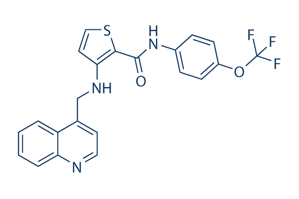
OSI-930 Chemical Structure
CAS No. 728033-96-3
Purity & Quality Control
Batch:
Purity:
99.92%
99.92
OSI-930 Related Products
| Related Products | Masitinib Amuvatinib (MP-470) Sitravatinib (MGCD516) ISCK03 PDGFR inhibitor 1 | Click to Expand |
|---|---|---|
| Related Compound Libraries | Kinase Inhibitor Library Tyrosine Kinase Inhibitor Library PI3K/Akt Inhibitor Library Cell Cycle compound library Angiogenesis Related compound Library | Click to Expand |
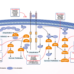
Signaling Pathway
Biological Activity
| Description | OSI-930 is a potent inhibitor of Kit (c-Kit), KDR and CSF-1R with IC50 of 80 nM, 9 nM and 15 nM, respectively; also potent to Flt-1, c-Raf and Lck and low activity against PDGFRα/β, Flt-3 and Abl. Phase 1. | |||||||||||
|---|---|---|---|---|---|---|---|---|---|---|---|---|
| Targets |
|
| In vitro | ||||
| In vitro | OSI-930 inhibits the cell proliferation in the HMC-1 cell line with IC50 of 14 nM without significant effect on growth of the COLO-205 cell line that does not express a constitutively active mutant receptor tyrosine kinase. Moreover, OSI-930 also induces apoptosis in HMC-1 cell line with EC50 of 34 nM. [1] A recent study shows that OSI-930 inactivates purified, recombinant cytochrome P450 (P450) 3A4 with a Ki of 24 μM in a time- and concentration-dependent mode. [2] | |||
|---|---|---|---|---|
| Kinase Assay | Protein kinase assays | |||
| Protein kinase assays are either done in-house by ELISA-based assay methods (Kit, KDR, PDGFRα, and PDGFRβ) or by a radiometric method. In-house ELISA assays used poly(Glu:Tyr) as the substrate bound to the surface of 96-well assay plates; phosphorylation is then detected using an antiphosphotyrosine antibody conjugated to HRP. The bound antibody is then quantitated using ABTS as the peroxidase substrate by measuring the absorbance at 405/490 nm. All assays uses purified recombinant kinase catalytic domains that are either expressed in insect cells or in bacteria. The Kit and EGFR protein used for in-house assays are prepared internally; other enzymes are obtained. Recombinant Kit protein is expressed as an NH2-terminal glutathione S-transferase fusion protein in insect cells and is initially purified as a nonphosphorylated (nonactivated) enzyme with a relatively high Km for ATP (400 μM). In some assays, an activated (tyrosine phosphorylated) form of the enzyme is prepared by incubation with 1 mM ATP for 1 hour at 30 °C. The phosphorylated protein is then passed through a desalting column to remove the majority of the ATP and stored at −80 °C in buffer containing 50% glycerol. The resultant preparation has a considerably higher specific activity and a lower Km for ATP (25 μM) than the initial nonphosphorylated preparation. The inhibition of Kit autophosphorylation by OSI-930 is assayed by incubation of the nonphosphorylated enzyme at 30 °C in the presence of 200 μM ATP and various concentrations of OSI-930. The reaction is stopped by removal of aliquots into SDS-PAGE sample buffer followed by heating to 100 °C for 5 minutes. The degree of phosphorylation of Kit is then determined by immunoblotting for both total Kit and phosphorylated Kit. | ||||
| Cell Research | Cell lines | HMC-1 and COLO-205 | ||
| Concentrations | 0--1 μM | |||
| Incubation Time | 48 hours | |||
| Method | For assays of cell proliferation and apoptosis, cells are seeded into 96-well plates and incubated for 2 to 3 days in the presence of OSI-930 at various concentrations. Inhibition of cell growth is determined by luminescent quantitation of the intracellular ATP content using CellTiterGlo. Induction of caspase-dependent apoptosis by OSI-930 is quantitated by an enzymatic caspase 3/7 assay. Inhibition of angiogenesis by OSI-930 is monitored using the rat aortic ring endothelial sprout outgrowth assay. Sections of aorta are prepared from CO2-euthanized male rats and cultured in vitro in a collagen matrix in the presence or absence of OSI-930. The collagen matrix is prepared from type 1 rat tail collagen solubilized in 0.1% acetic acid at 3 mg/mL, which is combined with 0.125 volume collagen buffer (0.05 N NaOH, 200 mM HEPES, 260 mM NaHCO3), 0.125 volume of medium 199, 0.0125 volume of 1 M NaOH, and 1% GlutaMax. Aortic rings are embedded in 0.4 mL of this matrix in six-well plates, to which 0.5 mL endothelial basal medium and the appropriate amount of OSI-930 is added; the rings are then incubated for 10 days and the resultant angiogenic sprout outgrowth is digitally quantitated from images by measurement of the sprout-containing area within a series of concentric rings around the aortic tissue area. | |||
| In Vivo | ||
| In vivo | OSI-930, administrated at the maximally efficacious dose of 200 mg/kg by oral gavage, exhibits potent antitumor activity in a broad range of preclinical xenograft models including HMC-1, NCI-SNU-5, COLO-205 and U251 xenograft models. [1] | |
|---|---|---|
| Animal Research | Animal Models | HMC-1, NCI-SNU-5, COLO-205 and U251 cells are injected s.c. into the right flank of CD1 nu/nu mice. |
| Dosages | ≤200 mg/kg | |
| Administration | Administered via p.o. | |
| NCT Number | Recruitment | Conditions | Sponsor/Collaborators | Start Date | Phases |
|---|---|---|---|---|---|
| NCT00513851 | Completed | Advanced Solid Tumors |
Astellas Pharma Inc|OSI Pharmaceuticals |
April 2006 | Phase 1 |
Chemical Information & Solubility
| Molecular Weight | 443.44 | Formula | C22H16F3N3O2S |
| CAS No. | 728033-96-3 | SDF | Download OSI-930 SDF |
| Smiles | C1=CC=C2C(=C1)C(=CC=N2)CNC3=C(SC=C3)C(=O)NC4=CC=C(C=C4)OC(F)(F)F | ||
| Storage (From the date of receipt) | |||
|
In vitro |
DMSO : 89 mg/mL ( (200.7 mM) Moisture-absorbing DMSO reduces solubility. Please use fresh DMSO.) Ethanol : 3 mg/mL Water : Insoluble |
Molecular Weight Calculator |
|
In vivo Add solvents to the product individually and in order. |
In vivo Formulation Calculator |
||||
Preparing Stock Solutions
Molarity Calculator
In vivo Formulation Calculator (Clear solution)
Step 1: Enter information below (Recommended: An additional animal making an allowance for loss during the experiment)
mg/kg
g
μL
Step 2: Enter the in vivo formulation (This is only the calculator, not formulation. Please contact us first if there is no in vivo formulation at the solubility Section.)
% DMSO
%
% Tween 80
% ddH2O
%DMSO
%
Calculation results:
Working concentration: mg/ml;
Method for preparing DMSO master liquid: mg drug pre-dissolved in μL DMSO ( Master liquid concentration mg/mL, Please contact us first if the concentration exceeds the DMSO solubility of the batch of drug. )
Method for preparing in vivo formulation: Take μL DMSO master liquid, next addμL PEG300, mix and clarify, next addμL Tween 80, mix and clarify, next add μL ddH2O, mix and clarify.
Method for preparing in vivo formulation: Take μL DMSO master liquid, next add μL Corn oil, mix and clarify.
Note: 1. Please make sure the liquid is clear before adding the next solvent.
2. Be sure to add the solvent(s) in order. You must ensure that the solution obtained, in the previous addition, is a clear solution before proceeding to add the next solvent. Physical methods such
as vortex, ultrasound or hot water bath can be used to aid dissolving.
Tech Support
Answers to questions you may have can be found in the inhibitor handling instructions. Topics include how to prepare stock solutions, how to store inhibitors, and issues that need special attention for cell-based assays and animal experiments.
Tel: +1-832-582-8158 Ext:3
If you have any other enquiries, please leave a message.
* Indicates a Required Field
Tags: buy OSI-930 | OSI-930 supplier | purchase OSI-930 | OSI-930 cost | OSI-930 manufacturer | order OSI-930 | OSI-930 distributor


