|
How to Cite 1. For In-Text Citation (Materials & Methods): 2. For Key Resources Table: |
||
|
Toll Free: (877) 796-6397 -- USA and Canada only -- |
Fax: +1-832-582-8590 Orders: +1-832-582-8158 |
Tech Support: +1-832-582-8158 Ext:3 Please provide your Order Number in the email. We strive to reply to |
Technical Data
| Formula | C24H24N6O2S3 |
||||||||||
| Molecular Weight | 524.68 | CAS No. | 314045-39-1 | ||||||||
| Solubility (25°C)* | In vitro | DMSO | 25 mg/mL (47.64 mM) | ||||||||
| Water | Insoluble | ||||||||||
| Ethanol | Insoluble | ||||||||||
| In vivo (Add solvents to the product individually and in order) |
|
||||||||||
|
* <1 mg/ml means slightly soluble or insoluble. * Please note that Selleck tests the solubility of all compounds in-house, and the actual solubility may differ slightly from published values. This is normal and is due to slight batch-to-batch variations. * Room temperature shipping (Stability testing shows this product can be shipped without any cooling measures.) |
|||||||||||
Preparing Stock Solutions
Biological Activity
| Description | BPTES is a potent and selective Glutaminase GLS1 (KGA) inhibitor with IC50 of 0.16 μM. It has no effect on glutamate dehydrogenase activity and causes only a very slight inhibition of γ-glutamyl transpeptidase activity. | ||
|---|---|---|---|
| Targets |
|
||
| In vitro | BPTES inhibits glutaminase activity expressed in human kidney cells with IC50 of 0.18 μM, and inhibits glutamate efflux by microglia with IC50 of 80-120 nM. [1] This compound preferentially slows cell growth in D54 cells with mutant IDH1. It also inhibits glutaminase activity, lowers glutamate and α-KG levels, and increases glycolytic intermediates. [2] This chemical (10 μM) inhibits cell growth of mHCC 3–4 cells derived from LAP/MYC tumors. It also inhibits growth of a MYC-dependent P493 cells by blocking DNA replication, leading to cell death and fragmentation. [3] |
||
| In vivo | In LAP/MYC mice, BPTES (12.5 mg/kg, i.p.) prolongs survival with no significant effects on MYC, GLS, or GLS2 levels. This compound (200 μg/mouse, i.p.) also inhibits tumor cell growth in mice harboring P493 tumor xenografts. [3] |
Protocol (from reference)
| Kinase Assay: |
|
|---|---|
| Cell Assay: |
|
| Animal Study: |
|
References
|
Customer Product Validation

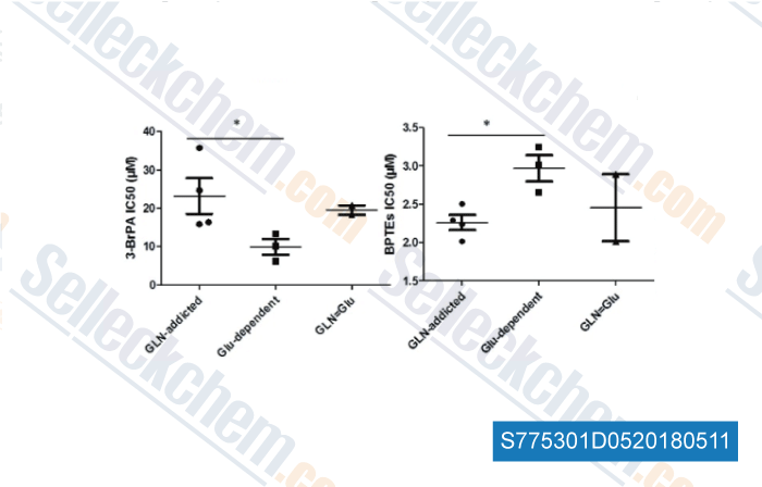
-
Data from [ , , Journal of Cancer, 2018, 9(9):1582-1591 ]

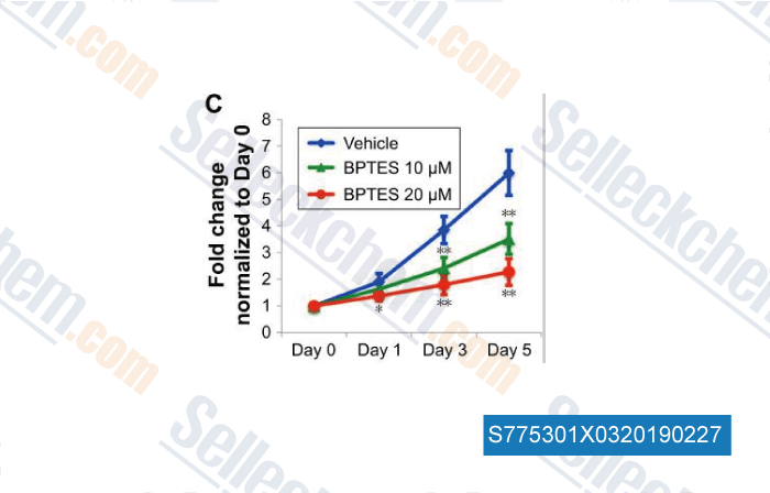
-
Data from [ , , Onco Targets Ther, 2018, 11:3721-3729 ]
Selleck's BPTES Has Been Cited by 63 Publications
| Identification of glutamine as a potential therapeutic target in dry eye disease [ Signal Transduct Target Ther, 2025, 10(1):27] | PubMed: 39837870 |
| Glutaminase inhibition confers antitumor effects in vitro and in vivo on tyrosine kinase inhibitors-resistant renal cell carcinoma cells by affecting vascular endothelial growth factor signaling [ Biomed Pharmacother, 2025, 191:118492] | PubMed: 40885145 |
| Live-cell metabolic analyzer protocol for measuring glucose and lactate metabolic changes in human cells [ STAR Protoc, 2025, 6(1):103518] | PubMed: 39799573 |
| Dual Inhibition of CDK4/6 and XPO1 Induces Senescence With Acquired Vulnerability to CRBN-Based PROTAC Drugs [ Gastroenterology, 2024, S0016-5085(24)00062-3] | PubMed: 38262581 |
| mTORC2-driven chromatin cGAS mediates chemoresistance through epigenetic reprogramming in colorectal cancer [ Nat Cell Biol, 2024, 10.1038/s41556-024-01473-0] | PubMed: 39080411 |
| Glutamine-derived aspartate is required for eIF5A hypusination-mediated translation of HIF-1α to induce the polarization of tumor-associated macrophages [ Exp Mol Med, 2024, 10.1038/s12276-024-01214-1] | PubMed: 38689086 |
| Glutaminolysis regulates endometrial fibrosis in intrauterine adhesion via modulating mitochondrial function [ Biol Res, 2024, 57(1):13] | PubMed: 38561846 |
| Targeting metabolic adaptive responses induced by glucose starvation inhibits cell proliferation and enhances cell death in osimertinib-resistant non-small cell lung cancer (NSCLC) cell lines [ Biochem Pharmacol, 2024, S0006-2952(24)00144-8] | PubMed: 38522556 |
| A novel method of Francisella infection of epithelial cells using HeLa cells expressing fc gamma receptor [ BMC Infect Dis, 2024, 24(1):1171] | PubMed: 39420255 |
| Glutamine metabolic microenvironment drives M2 macrophage polarization to mediate trastuzumab resistance in HER2-positive gastric cancer [ Cancer Commun (Lond), 2023, 43(8):909-937] | PubMed: 37434399 |
RETURN POLICY
Selleck Chemical’s Unconditional Return Policy ensures a smooth online shopping experience for our customers. If you are in any way unsatisfied with your purchase, you may return any item(s) within 7 days of receiving it. In the event of product quality issues, either protocol related or product related problems, you may return any item(s) within 365 days from the original purchase date. Please follow the instructions below when returning products.
SHIPPING AND STORAGE
Selleck products are transported at room temperature. If you receive the product at room temperature, please rest assured, the Selleck Quality Inspection Department has conducted experiments to verify that the normal temperature placement of one month will not affect the biological activity of powder products. After collecting, please store the product according to the requirements described in the datasheet. Most Selleck products are stable under the recommended conditions.
NOT FOR HUMAN, VETERINARY DIAGNOSTIC OR THERAPEUTIC USE.
